
Fig. S7
- ID
- ZDB-FIG-101008-10
- Publication
- Imai et al., 2010 - The ubiquitin proteasome system is required for cell proliferation of the lens epithelium and for differentiation of lens fiber cells in zebrafish
- Other Figures
- All Figure Page
- Back to All Figure Page
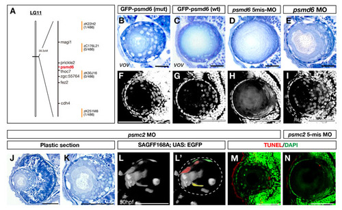
Lens phenotypes of vov mutants injected with GFP-tagged wild-type and vov mutant form psmd6 RNA and of psmd6 and psmc2 morphants. (A) The vov mutation is mapped in the genomic region between two polymorphic markers, zK22H2 and zK251M8, on zebrafish chromosome 11 (Vega Ver.32). Seven genes are annotated within this genomic region. (B,C) 72 hpf lens of vov mutant embryos injected with RNA encoding GFP-tagged Psmd6 carrying the vov mutation (B) and GFP-tagged wild-type Psmd6 (C). The introduction of wild-type psmd6 RNA inhibits the lens defects in the vov mutant, although some lens fiber nuclei are round in shape (C). (D,E) 72 hpf lens of the psmd6 five-mismatch morphant (D) and psmd6 morphant (E). The psmd6 morphant shows lens defects similar to the vov mutant. (F,G) DAPI labeling of 72 hpf lens of the vov mutant embryos injected with mRNA encoding GFP-tagged Psmd6 carrying the vov mutation (F) and GFP-tagged wild-type Psmd6 (G). The number of lens fiber nuclei positioned in the anterior half of the lens sphere is lower in vov mutant injected with wild-type psmd6 RNA than that of the vov-mutant form of psmd6 RNA. (H,I) DAPI labeling of 72 hpf lens of psmd6 five-mismatch morphant (H) and psmd6 morphant (I). Nuclei are swollen and retained in the anterior half of the lens sphere in the psmd6 morphant. (J,K) Plastic sections of 72 hpf psmc2 morphant eyes (J) and lens (K). Like the vov mutant, retinal apoptosis occurs (J) and round lens cell nuclei surround a small lens fiber core (K) in the psmc2 morphant lens. (L,L′) Confocal image of 50 hpf psmc2 morphant under the SAGFF168A; UAS:EGFP transgene. Lens fiber cells fail to fully elongate (pseudocolors in L′). (M,N) TUNEL of 72 hpf lens of psmc2 morphant (M) and psmc2 five-mismatch morphant (N). A few TUNEL signals (red) were detected in both cases. Nuclei were counterstained by DAPI (green). Scale bars: 50 μm. |