
Fig. S1
- ID
- ZDB-FIG-101007-49
- Publication
- Imai et al., 2010 - The ubiquitin proteasome system is required for cell proliferation of the lens epithelium and for differentiation of lens fiber cells in zebrafish
- Other Figures
- All Figure Page
- Back to All Figure Page
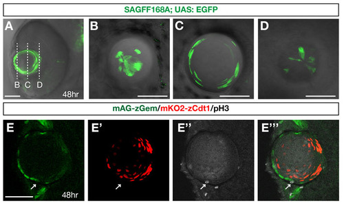
Zebrafish transgenic strains, SAGFF168A and Cecyil. (A) Ventral view of a zebrafish eye with the SAGFF168A; UAS:EGFP transgene at 48 hpf. GFP (green) is expressed in the lens. (B-D) Scanning of planes at the anterior-most surface (B), near the equator (C) and the posterior-most region of the lens sphere of 48 hpf SAGFF168A; UAS:EGFP transgenic embryo. GFP-positive lens fibers elongate to become very flat at the equatorial region (C) and extend their termini towards anterior and posterior poles, where tips of lens fiber processes converge (B,D). (E-E′′′) Confocal images of lens of Cecyil transgenic embryos with mAG-zGem (green), mKO2-zCdt1 (red) and anti-phosphorylated histone H3 antibody (gray, E′′). In the Cecyil transgenic line, an N-terminal 190 amino acid domain of zebrafish Cdt1 tagged with mKO2 (mKO2-zCdt1) (red, E′) and an N-terminal 100 amino acid domain of zebrafish Geminin tagged with mAG (mAG-zGem) (green, E) are expressed under the control of the EF1a promoter. mAG-zGem and mKO2-zCdt1 are expressed in lens epithelial cells and in postmitotic lens fiber differentiating cells, respectively. The anterior lens epithelial cells undergoing M phase do not express mAG-zGem or mKO2-zCdt1 (arrow). E′′′ is a merged image of E, E′ and E′′. Scale bars: 50 µm. |