
Fig. S4
- ID
- ZDB-FIG-101007-52
- Publication
- Imai et al., 2010 - The ubiquitin proteasome system is required for cell proliferation of the lens epithelium and for differentiation of lens fiber cells in zebrafish
- Other Figures
- All Figure Page
- Back to All Figure Page
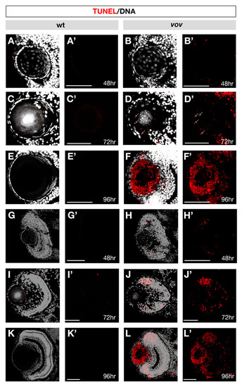
TUNEL in wild-type and the vov mutant lens and retina. (A-L) TUNEL (red) and DAPI (gray) staining of wild-type lens (A,C,E) and retina (G,I,K) and vov mutant lens (B,D,F) and retina (H,J,L) at 48 hpf (A,B,G,H), 72 hpf (C,D,I,J) and 96 hpf (E,F,K,L). (A′-L′) The same images as A-L shown through only a red channel (TUNEL). No TUNEL signal could be detected in the wild-type lens and retina at 48, 72 and 96 hpf and vov mutant lens at 48 hpf. However, in the vov mutant, small dotted signals are observed at 72 hpf, and most of the lens fiber cells surrounding a small lens fiber core are TUNEL positive, whereas the lens epithelium is TUNEL negative, suggesting that a majority of lens fiber cells undergo apoptosis-like degradation in the vov mutant at 96 hpf, except for a small central lens fiber core. In the vov mutant retina, TUNEL signals are observed in the neurogenic region near retinal CMZ at 48 hpf (H,H′). TUNEL signals increase at 72 hpf (J,J′) and 96 hpf (L,L′). Scale bars: 50 µm. |