
Fig. 2
- ID
- ZDB-FIG-100910-2
- Publication
- Coutelle et al., 2001 - Hedgehog signalling is required for maintenance of myf5 and myoD expression and timely terminal differentiation in zebrafish adaxial myogenesis
- Other Figures
- All Figure Page
- Back to All Figure Page
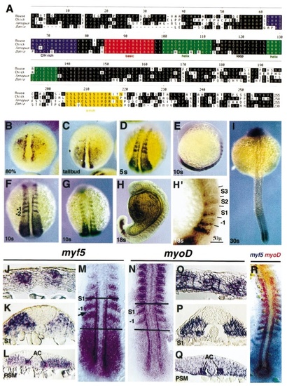
CLUSTAL alignment of the conceptual translation product of the zebrafish myf5 gene with representatives of other major vertebrate groups (mammals, birds, amphibia) (A). Localisation of myf5 (B–M) and myoD (N–Q) transcripts by in situ hybridisation at different developmental stages. Myf5 transcripts first appear in the presegmentation embryo in two stripes adjacent to the notochord and in broad triangular-shaped domains on each side of the embryonic shield at 9 hpf (80% epiboly) (B). The triangular expression domains appear to transit towards the posterior end in 10-hpf (tailbud stage) embryos (C). At the onset of segmentation lateral stripes of expression are present in the presomitic mesoderm and in the first 2–3 somites at 12 hpf (5– 6 somites); dorsal view (D). The maximal extent of myf5 expression, including the first 5– 6 somites, is reached at 14 hpf (10 –11 somites), including the lateral presomitic cells; lateral view (E), dorsal posterior view (F), dorsal anterior view (G). At subsequent stages, myf5 is rapidly down-regulated in rostral somites, 18 hpf (18 somites) (H, lateral view, and at higher magnification showing the location of myf5 expression in the posterior region of somites -I to IV, H′). Expression in the tail adjacent to the notochord is visible at 24 hpf (30 somites) (I). Flat-mounted dorsal view of 15-hpf (12–13 somite) embryo (M), showing down-regulation of myf5 in presomitic mesoderm in the region of somite -I (arrow). Sections from similar 15-hpf embryos: parasagittal in nascent somites (J, anterior to left), transverse through somite 1 (K), and transverse through presomitic mesoderm (L). Localisation of myoD: 15-hpf (12–13 somite) at mount (N) and sections: parasagittal (O, anterior to left), transverse through somite 1 (P), and transverse through presomitic mesoderm (Q). Both myf5 and myoD signals are most intensely reactive in adaxial cells in presomitic mesoderm and in lateral posterior cells of forming somites, in which myoD persists after myf5 declines. Neither myf5 nor myoD is highly expressed in the medial tail bud. Two-colour in situ mRNA hybridisation (R) with a red myoD probe and a purple myf5 probe at 13 hpf (7– 8 somites) dorsal view shows that myf5 and myoD are expressed in partially overlapping domains, in both the posterior somites and the adaxial cells. |
| Genes: | |
|---|---|
| Fish: | |
| Anatomical Terms: | |
| Stage Range: | 75%-epiboly to 26+ somites |
Reprinted from Developmental Biology, 236(1), Coutelle, O., Blagden, C.S., Hampson, R., Halai, C., Rigby, P.W.J., and Hughes, S.M., Hedgehog signalling is required for maintenance of myf5 and myoD expression and timely terminal differentiation in zebrafish adaxial myogenesis, 136-150, Copyright (2001) with permission from Elsevier. Full text @ Dev. Biol.