
Fig. 6
- ID
- ZDB-FIG-100910-6
- Publication
- Coutelle et al., 2001 - Hedgehog signalling is required for maintenance of myf5 and myoD expression and timely terminal differentiation in zebrafish adaxial myogenesis
- Other Figures
- All Figure Page
- Back to All Figure Page
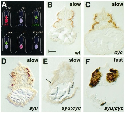
Exacerbation of slow muscle deficits in sonic you;cyclops double mutant. Schematic representation of hedgehog gene expression in wild-type and mutant zebrafish embryos (A). Slow MyHC-expressing muscle cells (B–E) in the yolk extension region of 24-hpf wild-type (B) and cyc (C) embryos are indistinguishable. syu embryos show slow muscle reduction (D). A syu;cyc double mutant embryo has only isolated residual slow muscle cells (E, arrow), yet expresses substantially normal fast muscle in an adjacent section (F). All panels show transverse sections, dorsal at top. Each of three syu;cyc mutants from 91 cross progeny, identified by lack of oorplate and U-shaped somites,were serially sectioned and individuals were found to contain two, five, and eight slow cells. Bar, 50 μm. |
| Antibodies: | |
|---|---|
| Fish: | |
| Anatomical Terms: | |
| Stage: | Prim-5 |
| Fish: | |
|---|---|
| Observed In: | |
| Stage: | Prim-5 |
Reprinted from Developmental Biology, 236(1), Coutelle, O., Blagden, C.S., Hampson, R., Halai, C., Rigby, P.W.J., and Hughes, S.M., Hedgehog signalling is required for maintenance of myf5 and myoD expression and timely terminal differentiation in zebrafish adaxial myogenesis, 136-150, Copyright (2001) with permission from Elsevier. Full text @ Dev. Biol.