
Fig. 5
- ID
- ZDB-FIG-100903-14
- Publication
- Jeong et al., 2010 - Inhibition of Plk1 induces mitotic infidelity and embryonic growth defects in developing zebrafish embryos
- Other Figures
- All Figure Page
- Back to All Figure Page
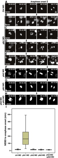
Live-cell imaging reveals that Plk1 is essential in mitotic progression, but Plk2 and Plk3 are dispensable in zebrafish embryos. (A–H) Transgenic zebrafish embryos expressing H2B-GFP were injected with 0.5 pmol of control (A), plk1 ATG MO (B-E), plk2 splicing MO (F), plk3 splicing MO (G), plk2 splicing MO and plk3 splicing MO (H, supplemental movie 8). The surface of the yolk was subjected to time-lapse microscopy, and images were captured every 2.5 min in 22 hpf embryos. The time from NEBD is shown as h:min:s. Black arrows mark the point of anaphase onset. White arrows mark the uncongressed chromosomes in B, lagging chromosomes in D, and multiple metaphase plates in E, respectively. Scale bar, 10 μm. (I) Box plots of mitotic timing, measured from NEBD to the onset of anaphase. At least 30 cells from more than five embryos each were scored. Bars within the boxes are the median values, as determined by statistical analysis using SPSS software. |
| Fish: | |
|---|---|
| Knockdown Reagents: | |
| Observed In: | |
| Stage: | 26+ somites |
Reprinted from Developmental Biology, 345(1), Jeong, K., Jeong, J.Y., Lee, H.O., Choi, E., and Lee, H., Inhibition of Plk1 induces mitotic infidelity and embryonic growth defects in developing zebrafish embryos, 34-48, Copyright (2010) with permission from Elsevier. Full text @ Dev. Biol.