
Fig. 2
- ID
- ZDB-FIG-100903-11
- Publication
- Jeong et al., 2010 - Inhibition of Plk1 induces mitotic infidelity and embryonic growth defects in developing zebrafish embryos
- Other Figures
- All Figure Page
- Back to All Figure Page
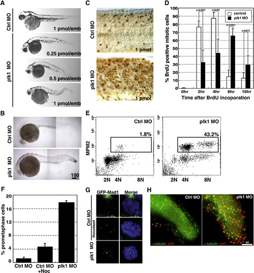
Depletion of Plk1 expression results in growth failure and apoptosis due to activation of SAC. (A) Zebrafish embryos were injected with various concentrations of control (Ctrl) or plk1 ATG MO (Plk1 MO) at the one- to four-cell stages. Photographs were taken at 48 hpf. (B) Embryos were injected with 0.5 pmol of control or plk1 ATG MO, and apoptosis was assayed by TUNEL staining at 28 hpf. Apoptotic cells are shown as purple dots. (C) Embryos were injected with 1 pmol of control or plk1 ATG MO and stained with anti-phosphohistone H3, Ser10 (pH3) antibodies at 24 hpf. (D) Time course from S phase into/out of G2/M, as shown by BrdU incorporation followed by double pH3/BrdU staining at indicated time points post BrdU pulse. Embryos injected with 0.25 pmol of control or plk1 ATG MO are compared. The percentage of BrdU/pH3 double-positive cells are represented in bar graphs (mean ± s.e.m.; n ≥ 385 cells from 2 embryos each). (E) MPM-2 staining for mitotic index measurement. X-axis, 7AAD for DNA staining; Y-axis, MPM-2 staining. (F) Comparison of the percentage of cells with GFP-Mad1 at the prometaphase kinetochores in control, nocodazole-treated, and plk1 MO-injected embryos. The results are the average of two independent experiments. At least 15 embryos each were analyzed in each experiment. (G) Localization of Mad1 at the prometaphase kinetochores in (F). Mad1 localization to kinetochores was not affected by the presence or absence of Plk1. (H) Embryos injected with 0.5 pmol of control or plk1 ATG MO were subjected to co-staining with anti-α-tubulin and anti-pH3 antibodies at 24 hpf. Optical sections were acquired every 1 μm at the end of the tails, merged and deconvoluted. Green, α-tubulin; red, pH3. Scale bar, 50 μm. Enlarged images are presented in Supplemental Fig. S2. |
| Fish: | |
|---|---|
| Knockdown Reagent: | |
| Observed In: | |
| Stage Range: | Prim-5 to Long-pec |
Reprinted from Developmental Biology, 345(1), Jeong, K., Jeong, J.Y., Lee, H.O., Choi, E., and Lee, H., Inhibition of Plk1 induces mitotic infidelity and embryonic growth defects in developing zebrafish embryos, 34-48, Copyright (2010) with permission from Elsevier. Full text @ Dev. Biol.