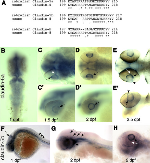
Claudin-5a and 5b expression in hyaloid vasculature. (A) The 3′ ends of three zebrafish claudin genes have significant homology with the C-terminal of mouse claudin-5. Claudin-5a (zgc 85723; GenBank: NM_213274) and claudin-5b (zgc 103419; GenBank: NM_001006044) are mostly homologous to claudin-5a of Fugu rubripes, with 82% identical (plus 8% similar) and 75% identical (plus 14% similar) amino acid sequences, respectively. The zebrafish claudin-h (GenBank: NM_131767) is mostly homologous to claudin-3a of Fugu. (B-E′) Whole mount in situ hybridization of claudin-5a expression. (B) Claudin-5a is expressed in the CNS (midbrain, hindbrain, ventricular zone, and epiphysis) at 1 dpf. (C-E & C′-E′) From 1.5 to 2.5 dpf, claudin-5mRNA is detected in the hyaloid vasculature (black arrows), hyaloid artery (white arrows) and the cornea (arrow heads). (F-H) Claudin-5b is expressed in the entire vascular system at 1 dpf (arrow and arrowheads in F), and is confined to the blood vessels of the brain (arrows in G) and cardiovascular system (arrowhead in G) at 2 dpf. Similar to claudin-5a, expression of claudin-5b in the hyaloid vasculature (black arrow in H) and hyaloid artery (white arrow in H) is seen at 1.5 dpf and lasts till 3 dpf. B-E, dorsal view; C′-E′, side view; F-H, side view.
|
