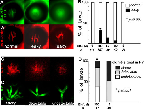
Bradykinin mediated disruption of zebrafish BRB (A, A′ & B). The l-fabp:DBP-EGFP;flk1:mCherry double transgenic larvae were treated with 8 to 100 μM BK from 5 to 9 dpf. (A) DBP-EGFP (green) in hyaloid vessels of control (left panel), leaky hyaloid vessels (middle and right panel); (A′) endothelial lining of blood vessels, mCherry (red). (B) At 9 dpf, the larvae were scored for the presence of leaky hyaloid vessels. Both Fisher′s test and chi-square test indicate that the BK treatment results in significantly increased numbers of larvae showing GFP leakage compared with those treated with control buffer (P < 0.001). (C) Claudin-5 expression (red) was evaluated by whole mount immunohistochemistry in Tg(flk1:EGFP) larvae exposed to 100 μM BK and scored as strong expression (left panel), detectable (middle panel) and undetectable (right panel); (C′) endothelial lining of blood vessels, flk1:EGFP (green). (D) Approximately 47 BK treated and 46 untreated flk1:EGFP larvae were evaluated for claudin-5 expression in the hyaloid vasculature.
|
