
FIGURE
Fig. 2
- ID
- ZDB-FIG-100809-21
- Publication
- Wang et al., 2010 - Morphological and molecular evidence for functional organization along the rostrocaudal axis of the adult zebrafish intestine
- Other Figures
- All Figure Page
- Back to All Figure Page
Fig. 2
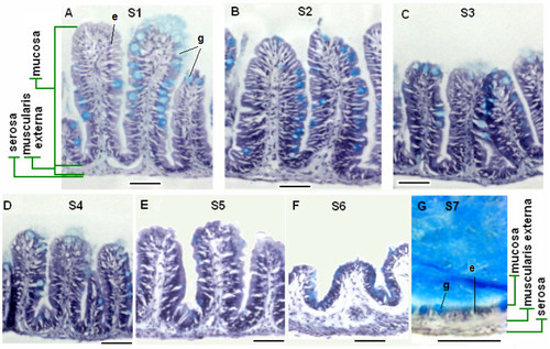
Histological features of adult zebrafish intestine along the seven anterior-posterior segments. (A-G) Representative cross sections of intestine from segments S1-S7 respectively. All sections were stained by Hematoxylin/Eosin/alcian blue. Segments S1-S6 contain three tissue layers: mucosa, muscularis externa and serosa, while S7 has a simple epithelium directly adjacent to the muscularis externa. Goblet cells (stained blue) are interspersed among the absorptive cells. Examples of enterocytes (e) and goblet cells (g) are indicated in Panels (A) and (G). Scale bars: 50 μm. |
Expression Data
Expression Detail
Antibody Labeling
Phenotype Data
Phenotype Detail
Acknowledgments
This image is the copyrighted work of the attributed author or publisher, and
ZFIN has permission only to display this image to its users.
Additional permissions should be obtained from the applicable author or publisher of the image.
Full text @ BMC Genomics