
- Title
-
Morphological and molecular evidence for functional organization along the rostrocaudal axis of the adult zebrafish intestine
- Authors
- Wang, Z., Du, J., Lam, S.H., Mathavan, S., Matsudaira, P., and Gong, Z.
- Source
- Full text @ BMC Genomics
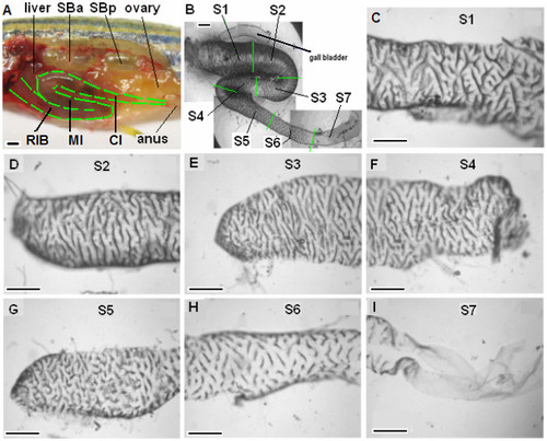
Anatomical features of adult zebrafish intestine. (A) A partially dissected 6-month-old zebrafish to show the folding of the three portion of intestine in vivo: rostral intestinal bulb (RIB), mid-intestine (MI) and caudal intestine (CI). Liver, ovary, anus, swimbladder anterior (SBa) and posterior (SBp) chambers are indicated. (B) An isolated zebrafish intestine in vitro after removal of the surrounding mesentery. The isolated intestine was divided into seven roughly equal-length segments as indicated by green lines: S1-S2 from RIB, S3-S4 from MI and S5-S7 from CI. The associated gall bladder is indicated. (C - I) Surface views of segments S1-S7 showing the folding of the mucosal surface into circumferential ridges. Scale bars, 500 μm. |
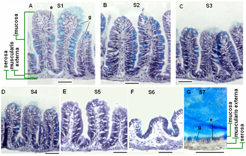
Histological features of adult zebrafish intestine along the seven anterior-posterior segments. (A-G) Representative cross sections of intestine from segments S1-S7 respectively. All sections were stained by Hematoxylin/Eosin/alcian blue. Segments S1-S6 contain three tissue layers: mucosa, muscularis externa and serosa, while S7 has a simple epithelium directly adjacent to the muscularis externa. Goblet cells (stained blue) are interspersed among the absorptive cells. Examples of enterocytes (e) and goblet cells (g) are indicated in Panels (A) and (G). Scale bars: 50 μm. |

ZFIN is incorporating published figure images and captions as part of an ongoing project. Figures from some publications have not yet been curated, or are not available for display because of copyright restrictions. |
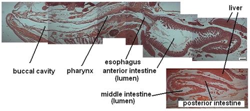
Anatomy of adult zebrafish showing the digestive tract. A composite of H&E sections from the medial-longitudinal plane of a male zebrafish reveals the main components of the digestive tract. Scale bar, 500 μm. |
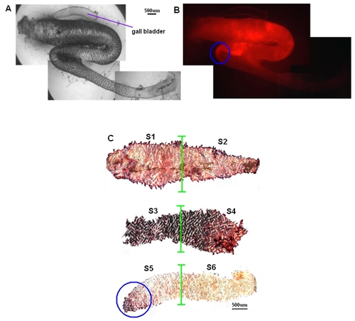
Expression of fabp2 gene in adult zebrafish intestine. (A) Isolation of an intestine from a Tg(fabp2:RFP) fish. (B) Expression of fabp2 in a Tg(fabp2:RFP) fish as indicated by the RFP reporter. Circle, the junction where expression of fabp2:rfp transgene disappears. (C) In situ hybridization detection of endogenous fabp2 expression in adult zebrafish intestine from segment S1∼S7, respectively. High expression level is observed in segments S1-S4, but it is turn off nearby the second natural turn of the intestine (circle). Beyond this region, the expression level becomes undetectable. |