
Fig. 1
- ID
- ZDB-FIG-100809-20
- Publication
- Wang et al., 2010 - Morphological and molecular evidence for functional organization along the rostrocaudal axis of the adult zebrafish intestine
- Other Figures
- All Figure Page
- Back to All Figure Page
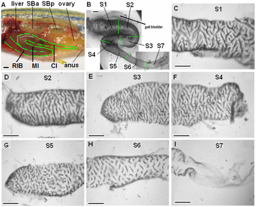
Anatomical features of adult zebrafish intestine. (A) A partially dissected 6-month-old zebrafish to show the folding of the three portion of intestine in vivo: rostral intestinal bulb (RIB), mid-intestine (MI) and caudal intestine (CI). Liver, ovary, anus, swimbladder anterior (SBa) and posterior (SBp) chambers are indicated. (B) An isolated zebrafish intestine in vitro after removal of the surrounding mesentery. The isolated intestine was divided into seven roughly equal-length segments as indicated by green lines: S1-S2 from RIB, S3-S4 from MI and S5-S7 from CI. The associated gall bladder is indicated. (C - I) Surface views of segments S1-S7 showing the folding of the mucosal surface into circumferential ridges. Scale bars, 500 μm. |