
FIGURE
Fig. 4
- ID
- ZDB-FIG-100628-65
- Publication
- Kim et al., 2010 - Gli2a protein localization reveals a role for Iguana/DZIP1 in primary ciliogenesis and a dependence of Hedgehog signal transduction on primary cilia in the zebrafish
- Other Figures
- All Figure Page
- Back to All Figure Page
Fig. 4
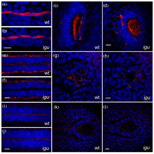
Motile cilia are largely unaffected by the loss of Iguana (Igu) function. Motile cilia (red: acetylated tubulin in a-h) in the pronephros at 48 hpf in igu mutant embryos appeared normal (b), as did those in the olfactory pits (d). By contrast motile cilia were largely absent from the floorplate at 28 hpf (f) and those in Kupffer′s vesicle were significantly reduced in number (h) in 10 somite stage igu mutant embryos. Basal bodies (red: γ-tubulin in i-l) in the igu mutants, by contrast, were formed normally at all stages examined in the floorplate (j) and Kupffer′s vesicle (l). Scale bars: 10 μm |
Expression Data
Expression Detail
Antibody Labeling
Phenotype Data
| Fish: | |
|---|---|
| Observed In: | |
| Stage Range: | 10-13 somites to Long-pec |
Phenotype Detail
Acknowledgments
This image is the copyrighted work of the attributed author or publisher, and
ZFIN has permission only to display this image to its users.
Additional permissions should be obtained from the applicable author or publisher of the image.
Open Access.
Full text @ BMC Biol.