
|
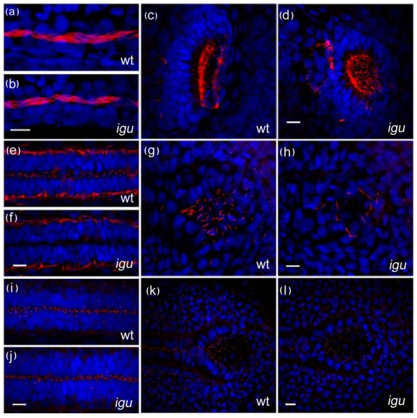
Fig. 4 Motile cilia are largely unaffected by the loss of Iguana (Igu) function. Motile cilia (red: acetylated tubulin in a-h) in the pronephros at 48 hpf in igu mutant embryos appeared normal (b), as did those in the olfactory pits (d). By contrast motile cilia were largely absent from the floorplate at 28 hpf (f) and those in Kupffer′s vesicle were significantly reduced in number (h) in 10 somite stage igu mutant embryos. Basal bodies (red: γ-tubulin in i-l) in the igu mutants, by contrast, were formed normally at all stages examined in the floorplate (j) and Kupffer′s vesicle (l). Scale bars: 10 μm