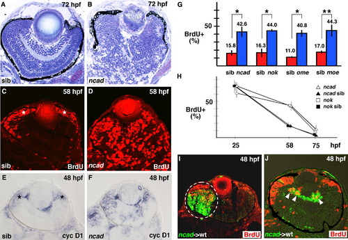
Ratio of number of proliferating cells to total number of retinal cells increases in zebrafish cell-polarity-defective mutants. (A and B) Plastic sections of 72 hpf wild-type retina (A) and ncad mutant retina (B). The laminar structure is severely disorganized in the ncad mutant retina. (C and D) BrdU labeling of 58 hpf wild-type retina (C) and ncad mutant retina (D). BrdU-positive cells (red) are located in CMZ of wild-type retinas (C, asterisks), whereas many retinal cells are BrdU-positive even in the central region of ncad mutant retinas (D). (E and F) In situ hybridization of 48 hpf wild-type retina (E) and ncad mutant retina (F) using cyclin D1 RNA probe. cyclin D1 is expressed in CMZ of wild-type retinas (E, asterisks), whereas it is expressed in not only CMZ but also the central region of ncad mutant retinas (F). (G) Percentage of BrdU-positive cells with respect to total number of retinal cells at 58 hpf in retinal-cell-polarity-defective mutants: ncad, nok, ome, and moe (blue bars) and their wild-type siblings (red bars). The ratio of BrdU-positive cells is 2.7 times higher in ncad, nok, and moe mutants and 3.7 times higher in the ome mutant than in their siblings. Student’s t-test; *p < 0.005 and **p < 0.01. (H) Temporal profile of the percentage of BrdU-positive cells with respect to total number of retinal cells in nok (open squares) and ncad (open triangles) embryos and their wild-type sibling embryos (filled squares and triangles). The percentage is higher in these mutants than in wild-type siblings at 58 hpf, but decreased to the wild-type level by 75 hpf. The difference between the ncad/nok mutants and their siblings is not significant (Student’s t-test, p > 0.05) at both 25 and 75 hpf, but significant (Student’s t-test p < 0.05) at 58 hpf. (I and J) Cell-transplantation experiments of ncad mutant donor cells into wild-type recipient retinas. A large clone of mutant cells forms very close to CMZ of the wild-type retina (I) and a small clone forms in the vitrial region of the neural retina (J). In both cases, ncad mutant cells (green) abnormally incorporate BrdU (red) in wild-type recipient retinas [white dashed circle (I) and arrowheads (J)].
|
