
Fig. 5
- ID
- ZDB-FIG-100506-27
- Publication
- Williams et al., 2010 - Hedgehog signaling induces arterial endothelial cell formation by repressing venous cell fate
- Other Figures
- All Figure Page
- Back to All Figure Page
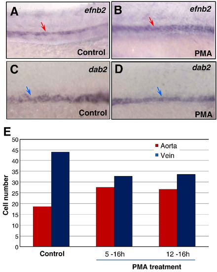
Activated Hh signaling causes an increase of arterial cells and loss of venous cells. (A–D) In situ hybridization analysis displaying the increased efnb2 expression in purmorphamine (PMA)-treated embryos (A) compared to controls (B); and the reduced dab2 expression in embryos treated with PMA (C) compared to controls (0.1% DMSO) (D). (E) Bar chart depicting numbers of arterial and venous cells in PMA-treated embryos from 5 to 16 hpf and 12 to 16 hpf, compared to controls (0.1% DMSO). Arterial cell number: 19.3 ± 0.6 (control); 27.5 ± 0.9 (5–16 hpf; PMA treated); 26.7 ± 0.6 (12–16 hpf; PMA treated). Venous cell number: 41.6 ± 1.5 (control); 32.7 ± 1.2 (5–16 hpf; PMA treated); 33.5 ± 1.8 (12–16 hpf; PMA treated). Endothelial cells were counted from 6 sections derived from 3 wild-type embryos or 3 PMA-treated embryos. Each section covers four segment lengths of intersomitic vessels along the dorsal aorta and the posterior cardinal vein in the middle trunk above the yolk extension region. a: aorta. v: vein. |
Reprinted from Developmental Biology, 341(1), Williams, C., Kim, S.H., Ni, T.T., Mitchell, L., Ro, H., Penn, J.S., Baldwin, S.H., Solnica-Krezel, L., and Zhong, T.P., Hedgehog signaling induces arterial endothelial cell formation by repressing venous cell fate, 196-204, Copyright (2010) with permission from Elsevier. Full text @ Dev. Biol.