
Fig. 1
- ID
- ZDB-FIG-100506-23
- Publication
- Williams et al., 2010 - Hedgehog signaling induces arterial endothelial cell formation by repressing venous cell fate
- Other Figures
- All Figure Page
- Back to All Figure Page
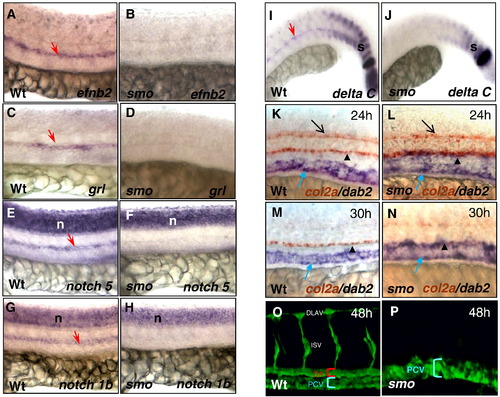
smo mutants display expansion of the posterior cardinal vein and absence of the dorsal aorta. (A–J) Lateral views displaying absent expression of efnb2 (A, B), grl (C, D), notch5 (E, F), notch1b (G, H) and deltaC (I, J) in smo mutants, compared to wild-type embryos. (K–N) Double in situ hybridization exhibiting the expansion of dab2 venous domain and col2a expression in the neural tube and the hypocord in smo mutants (L, N) compared to wild-type embryos (K, M). (O, P) Confocal microscopy depicting the dorsal aorta and the posterior cardinal vein in wild-type embryos [Tg(flk:EGFP)] (O); and absence of the dorsal aorta and expansion of the posterior cardinal veins in smo mutants [smohi640/smohi640; Tg(flk:EGFP)] (P). Red arrow: dorsal aorta. Blue arrow: cardinal vein. Black arrow: floor plate. Black arrowhead: hypocord. s: somite. n: neural tube. DA: dorsal aorta. PCV: posterior cardinal vein. DLAV: dorsal longitudinal anastomotic vessel. |
Reprinted from Developmental Biology, 341(1), Williams, C., Kim, S.H., Ni, T.T., Mitchell, L., Ro, H., Penn, J.S., Baldwin, S.H., Solnica-Krezel, L., and Zhong, T.P., Hedgehog signaling induces arterial endothelial cell formation by repressing venous cell fate, 196-204, Copyright (2010) with permission from Elsevier. Full text @ Dev. Biol.