
|
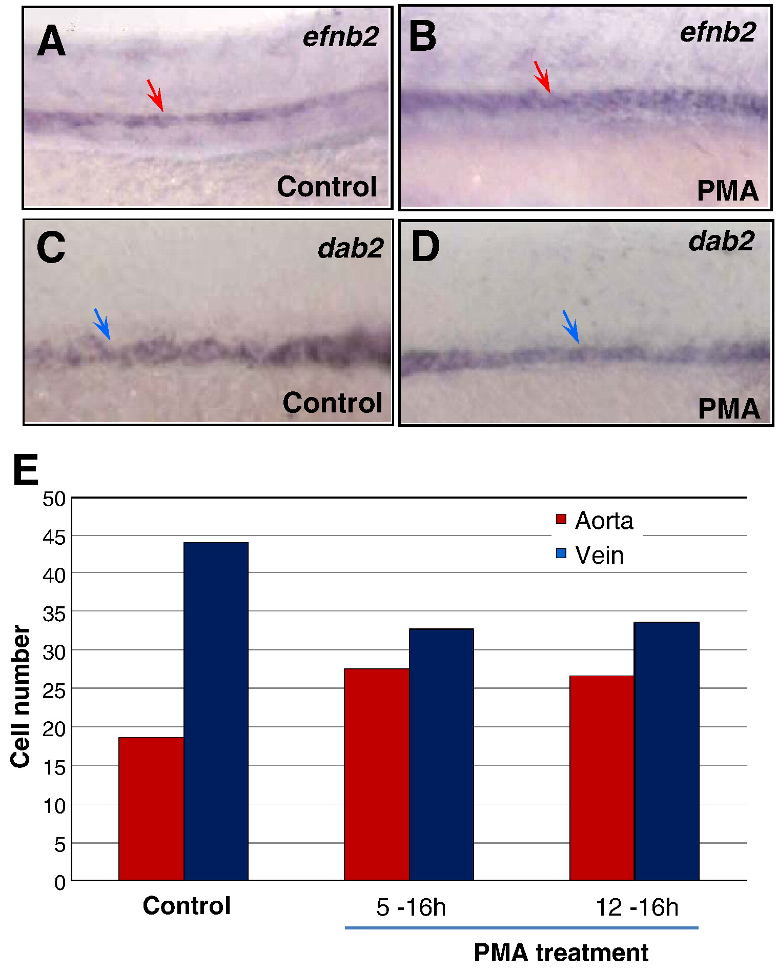
Fig. 5 Activated Hh signaling causes an increase of arterial cells and loss of venous cells. (A–D) In situ hybridization analysis displaying the increased efnb2 expression in purmorphamine (PMA)-treated embryos (A) compared to controls (B); and the reduced dab2 expression in embryos treated with PMA (C) compared to controls (0.1% DMSO) (D). (E) Bar chart depicting numbers of arterial and venous cells in PMA-treated embryos from 5 to 16 hpf and 12 to 16 hpf, compared to controls (0.1% DMSO). Arterial cell number: 19.3 ± 0.6 (control); 27.5 ± 0.9 (5–16 hpf; PMA treated); 26.7 ± 0.6 (12–16 hpf; PMA treated). Venous cell number: 41.6 ± 1.5 (control); 32.7 ± 1.2 (5–16 hpf; PMA treated); 33.5 ± 1.8 (12–16 hpf; PMA treated). Endothelial cells were counted from 6 sections derived from 3 wild-type embryos or 3 PMA-treated embryos. Each section covers four segment lengths of intersomitic vessels along the dorsal aorta and the posterior cardinal vein in the middle trunk above the yolk extension region. a: aorta. v: vein.
Reprinted from Developmental Biology, 341(1), Williams, C., Kim, S.H., Ni, T.T., Mitchell, L., Ro, H., Penn, J.S., Baldwin, S.H., Solnica-Krezel, L., and Zhong, T.P., Hedgehog signaling induces arterial endothelial cell formation by repressing venous cell fate, 196-204, Copyright (2010) with permission from Elsevier. Full text @ Dev. Biol.