
Fig. S3
- ID
- ZDB-FIG-100429-60
- Publication
- Pillay et al., 2010 - The Hox cofactors Meis1 and Pbx act upstream of gata1 to regulate primitive hematopoiesis
- Other Figures
- All Figure Page
- Back to All Figure Page
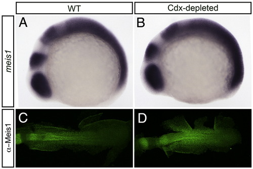
Cdx-depleted embryos exhibit increased levels of Meis1. (A, B) Shown are representative embryos following in situ hybridization analysis of meis1 expression in wild type (WT; A) compared with Cdx-depleted (B) embryos. The PLM of 14 hpf flat-mounted, deyolked embryos is shown in dorsal view with anterior to left. meis1 expression is increased in the posterior of Cdx-depleted embryos (B) when compared to WT (A). (C, D) Shown are representative embryos following immunohistochemical staining with the P2A6 antibody to visualize Meis1 protein levels in 14 hpf wild type (C) and Cdx-depleted (D) embryos. Flat-mount; dorsal view of embryo; anterior to left. Embryos are visualized under a 10x objective. Meis1 antibody staining is increased in Cdx-depleted embryos (D) when compared to WT (C). |
Reprinted from Developmental Biology, 340(2), Pillay, L.M., Forrester, A.M., Erickson, T., Berman, J.N., and Waskiewicz, A.J., The Hox cofactors Meis1 and Pbx act upstream of gata1 to regulate primitive hematopoiesis, 306-317, Copyright (2010) with permission from Elsevier. Full text @ Dev. Biol.