
Fig. 8
- ID
- ZDB-FIG-100429-56
- Publication
- Pillay et al., 2010 - The Hox cofactors Meis1 and Pbx act upstream of gata1 to regulate primitive hematopoiesis
- Other Figures
- All Figure Page
- Back to All Figure Page
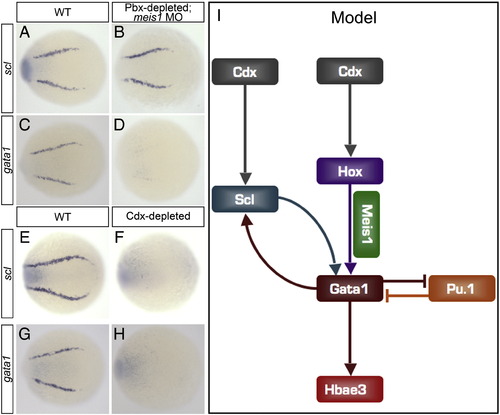
Unlike Cdx, Meis1 and Pbx function downstream of scl to activate gata1 expression. (A–H) Shown are representative embryos following in situ hybridization analysis of scl and gata1 expression in 12 hpf embryos. Dorsal view of gene expression in the posterior lateral-plate mesoderm (PLM) is shown in whole-mount embryos with anterior oriented to the left. Pbx-depleted; meis1-morphant embryos exhibit wild type (WT) levels of scl expression (B; 100%, n = 13) and abolished gata1 expression (D; 100%, n = 18) when compared to WT (A, C) embryos. Genotype of Pbx-depleted; meis1-morphant embryos was determined by in situ hybridization analysis of egr2b expression. Cdx-depleted embryos exhibit abolished scl (F; 100%, n = 5) and gata1 (H; 100%, n = 7) expression when compared to WT (E, G) embryos. (I) Hierarchical model indicating the genetic interactions that occur between a subset of transcription factors that regulate zebrafish primitive hematopoiesis. Arrows do not necessarily represent direct interactions. |
Reprinted from Developmental Biology, 340(2), Pillay, L.M., Forrester, A.M., Erickson, T., Berman, J.N., and Waskiewicz, A.J., The Hox cofactors Meis1 and Pbx act upstream of gata1 to regulate primitive hematopoiesis, 306-317, Copyright (2010) with permission from Elsevier. Full text @ Dev. Biol.