
Fig. 3
- ID
- ZDB-FIG-100429-51
- Publication
- Pillay et al., 2010 - The Hox cofactors Meis1 and Pbx act upstream of gata1 to regulate primitive hematopoiesis
- Other Figures
- All Figure Page
- Back to All Figure Page
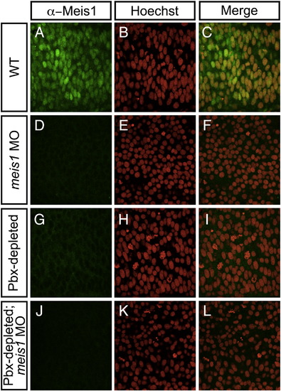
Meis1 protein levels are severely diminished in the posterior mesoderm of Pbx-depleted and meis1-morphant embryos. Shown are representative embryos following immunohistochemical staining with the P2A6 antibody (green; A, D, G, J) to visualize Meis1 protein levels, and Hoechst 33258 (red; B, E, H, K) to visualize nuclei in 14 hpf wild type (WT; A–C), meis1-morphant (D–F), Pbx-depleted (G–I), and Pbx-depleted; meis1-morphant (J–L) embryos. Flat-mount; dorsal view of posterior mesoderm; anterior to left. All embryos visualized under a 100x objective. In wild type (WT; A–C) embryos, α-Meis1 antibody staining is punctate, and colocalizes with Hoechst nuclear staining. meis1-morphant (D) and Pbx-depleted (G) embryos exhibit severely decreased α-Meis1 antibody staining when compared to WT embryos (A). α-Meis1 antibody staining is further abolished in Pbx-depleted; meis1-morphant embryos (J). α-Meis1 antibody staining does not colocalize with Hoechst nuclear staining in Pbx-depleted embryos (G–I). |
Reprinted from Developmental Biology, 340(2), Pillay, L.M., Forrester, A.M., Erickson, T., Berman, J.N., and Waskiewicz, A.J., The Hox cofactors Meis1 and Pbx act upstream of gata1 to regulate primitive hematopoiesis, 306-317, Copyright (2010) with permission from Elsevier. Full text @ Dev. Biol.