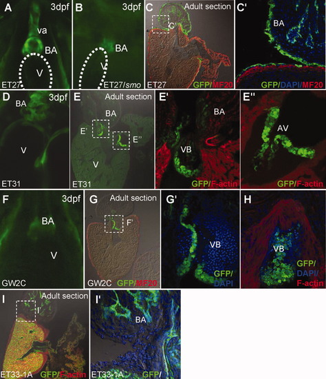
Developmental changes in CET lines with early BA-specific expression. A: EGFP expression in the BA and ventral aorta (VA) in wildtype 3-dpf ET27. B: smo mutation affects the ventral aorta, but not the domain of EGFP expression corresponding to BA. C, C′: ET27 adult heart sections shown at low (C) and high (C′) magnification. D: ET31 embryonic expression in BA. E, E′, E″: Cardiac valves; VB (E′) and AV (E″) marked in adult heart of ET31 shown on F-actin-stained sections. F: BA restricted embryonic expression of GW2C. G: GW2C adult heart sections at low magnification. G′, H: Serial sections of VB valve showing its different morphology at high magnification. I, I′: ET33-1A adult heart sections shown at low (I) and high (I″) magnification, for comparison of endocardal cell morphology in BA with ET27 (C′). A, B, D, F: Ventral view at 3dpf; C, C′: Sections of the adult heart immunostained for EGFP and MF20. E′, E″, H, I: Sections counterstained with F-actin. A-V, atrio-ventricular valve; BA, bulbus arteriosus; VB, ventriculo-bulbar valve; V, ventricle.
|
