
Fig. S3
- ID
- ZDB-FIG-100302-9
- Publication
- Li et al., 2010 - Regulation of neural crest cell fate by the retinoic acid and Pparg signalling pathways
- Other Figures
- All Figure Page
- Back to All Figure Page
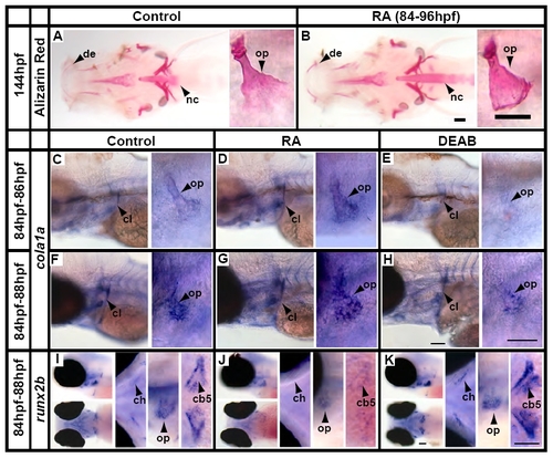
RA signalling simultaneously increases bone matrix synthesis while inhibiting runx2b expression. (A,B) Alizarin Red staining at 144 hpf shows that a late pulse treatment with RA results in increased ossification, particularly in the dentary (de), opercle (op) and notochord (nc). Low-magnification ventral views are shown to the left and high-magnification images of the opercle are shown to the right. (C-H) col1a1 is expressed around the developing bony skeleton including the cleithrum (cl) and opercle in untreated fish (C,F). This expression is increased after 2 hours (D) and 4 hours (G) of RA treatment and decreased by similar treatment with DEAB (E,H). Low-magnification side views are shown to the left and high-magnification images of the opercle are shown to the right. (I-K) runx2b is expressed around the developing bony skeleton, including the ceratohyal (ch), fifth ceratobranchial and pharyngeal teeth (cb5) and opercle in untreated fish (I). This expression is diminished by RA treatment (J), and is increased by DEAB treatment (K). Each panel shows low-magnification side and ventral views on the left and high-magnification images of the ceratohyal, fifth ceratobranchial and pharyngeal teeth and opercle on the right. Scale bars: 25 μm. |