
Fig. 1
- ID
- ZDB-FIG-100302-3
- Publication
- Li et al., 2010 - Regulation of neural crest cell fate by the retinoic acid and Pparg signalling pathways
- Other Figures
- All Figure Page
- Back to All Figure Page
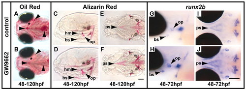
The Pparg inhibitor GW9662 blocks adipocyte formation while enhancing osteoblast differentiation. (A,B) Ventral views of Oil Red staining showing that lipid droplet accumulation throughout the zebrafish embryo head and cardiac region is strongly diminished by GW9662 treatment for 72 hours. Droplet accumulation (arrowheads) is highest at either end of the ceratohyal cartilage element (asterisk) and around the heart. (C-F) Alizarin Red staining shows that ossification is enhanced after GW9662 treatment for 72 hours. Dermal bones (bs, branchiostegal ray 3; ps, parasphenoid; op, opercle) and a cartilage bone (hm, hyomandibula) show increased levels of ossification. Side views (C,D) and ventral views (E,F) are shown. The eyes have been removed for clarity. Quantification for this experiment is shown in Table S1 in the supplementary material. (G-J) runx2b expression is strongly upregulated after 24 hours of GW9662 treatment. The opercle, branchiostegal ray 3 and the parasphenoid primordia all show more intense staining (H,J), consistent with the increase in ossification seen by 120 hpf (D,F). Scale bars: 100 μm in F for A-F and in J for G-J. |