
Fig. 2
- ID
- ZDB-FIG-100302-4
- Publication
- Li et al., 2010 - Regulation of neural crest cell fate by the retinoic acid and Pparg signalling pathways
- Other Figures
- All Figure Page
- Back to All Figure Page
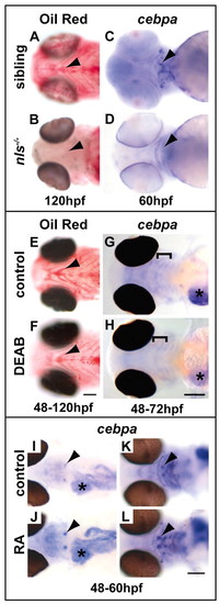
RA signalling is required for adipocyte differentiation. (A,B) Ventral views showing that lipid droplet accumulation throughout the head and cardiac region is lost in nls-/- fish at 120 hpf. Arrowheads point to droplets present in cells near to the distal end of the ceratohyal cartilage that are present in wild-type (A) and absent in nls-/- (B) fish. (C,D) Ventral views showing that expression of the adipocyte differentiation marker cepba is strongly reduced in nls-/- fish at 60 hpf. Arrowheads point to a cluster of cepba-positive cells at the distal end of the ceratohyal cartilage (C) that is lost in nls-/- embryos (D). Arrowheads point to droplets present in cells near to the distal end of the ceratohyal cartilage that are present in wild-type (C) and absent in nls-/- (D) fish. (E,F) Ventral views showing that lipid droplet accumulation throughout the head and cardiac region is reduced after treatment with DEAB. Arrowheads point to droplets present in cells near to the distal end of the ceratohyal cartilage element (E) that are reduced in DEAB-treated fish (F). (G,H) Dorsal views showing that cebpa expression throughout the head and liver is reduced after treatment with DEAB. Brackets indicate the area of diffuse expression in the pharyngeal region (G) that is reduced in treated fish (H). The liver also shows a strong reduction in cebpa expression levels after DEAB treatment (asterisk). (I-L) Retinoic acid (RA) treatment upregulates expression of cebpa after 12 hours. Dorsal flat-mounted fish showing that cebpa expression is particularly enhanced in two clusters of cells on either side of the heart (arrowheads in I and J) and liver (asterisk). Ventral views show enhanced expression in the pharyngeal arches, especially at the distal ends of the ceratohyal cartilage (arrowheads in K and L). Scale bars: 100 μm. |