
Fig. 4
- ID
- ZDB-FIG-091207-4
- Publication
- Furutani-Seiki et al., 1996 - Neural degeneration mutants in the zebrafish, Danio rerio
- Other Figures
- All Figure Page
- Back to All Figure Page
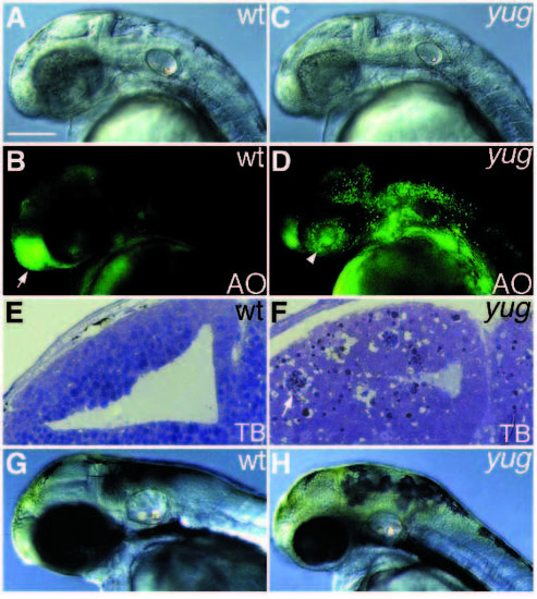
Class III mutant phenotypes. (A,B,E,G) Wild type; (C,D,F,H) yug embryos. (A-F) at 36 hours, (G,H) at 60 hours. (B,D) Acridine orange staining of A and C, respectively. (E,F) Parasagittal section of the brain. (A,C) The brain appears normal except for the presence of degenerating cells in yug embryos. (B,D) Apoptotic cells exist throughout the brain including the neural retina (arrowhead); acridine orange (AO)-staining of the olfactory epithelium (arrow) is reduced in yug; toluidine blue stained apoptotic cells are seen in the tectum in yug. The apoptotic cells are in a clump (arrow). Viable cells in the tectum are less well stained in yug embryos compared to wild type. The neuroepithelium is not differentiated properly (E,F). The head and eyes are small in yug embryos (G,H). Scale bar, 100 µm. |
| Fish: | |
|---|---|
| Observed In: | |
| Stage Range: | Prim-25 to Pec-fin |