
Fig. 5
- ID
- ZDB-FIG-091116-4
- Publication
- Furutani-Seiki et al., 1996 - Neural degeneration mutants in the zebrafish, Danio rerio
- Other Figures
- All Figure Page
- Back to All Figure Page
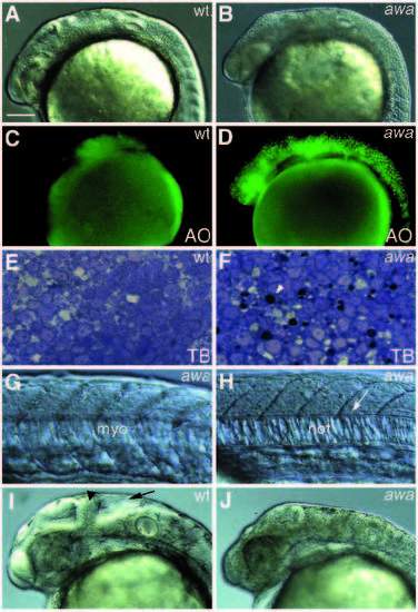
Class IV mutant phenotypes. (A,C,E,I) Wild type; (B,D,F,G,H,J) awa embryos. (A-H) at the 25-somite stage; (I,J) at 36 hours. (C,D) Acridine orange (AO)-staining of A and B, respectively. (E,F) Parasagittal section of the brain. (A-D) General apoptosis of neuroepithelium in the nervous system of awa; (E,F) toluidine blue (TB)-stained apoptotic cells (arrowhead) in awa. The rest of the cells in the neuroepithelium appear unaffected; (G,H) myotomes (myo), floor plate cells (arrow) and vacuolated cells of notochord (not) are relatively normal in awa embryos; (I,J) small and distorted head of awa embryos. The midbrain-hindbrain boundary (arrowhead), tectum and the fourth ventricle (arrow) are not well developed in awa embryos. Scale bar, 100 µm. |
| Fish: | |
|---|---|
| Observed In: | |
| Stage Range: | 20-25 somites to Prim-25 |