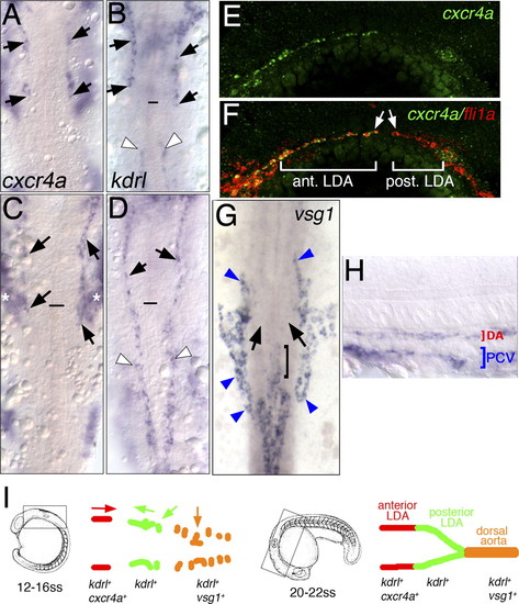
LDA progenitors are genetically distinct. (A–H) In situ hybridization with indicated markers. (A–D,G) Dorsal view, anterior is up. (E,F,H) Lateral view, anterior to the left, dorsal is up. (A–D) Expression of cxcr4a (A,C) and kdrl (B,D) at the 18-somite stage and the 22-somite stage, respectively. Anterior LDA (black arrows) and posterior LDA (white arrowheads) are indicated. Black line marks the anterior end of the notochord. Asterisks in C mark expression of cxcr4a in pharyngeal arch tissue. (E,F) Confocal images of 22-somite-stage embryos following double-fluorescent in situ hybridization. Endothelial cells are labeled by expression of fli1a in red and cxcr4a in green. (G,H) Expression of vsg1 at the 22-somite stage. (G) Dorsal view. (H) Lateral view. (G) Black arrows mark posterior LDA, which does not express vsg1. Bracket marks anterior end of DA. Blue arrowheads mark venous cells. (I) Schematic drawing of DA domains delineated by different migratory behaviors and gene expression profiles at the 18-somite stage and the 22-somite stage.
|
