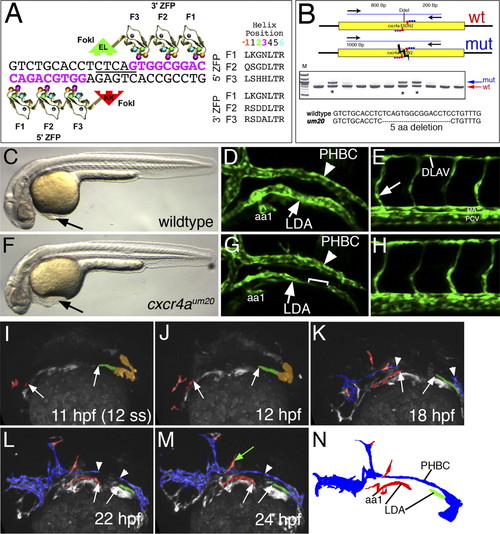
LDA formation defects in cxcr4a-deficient embryos. (A) Schematic depicting ZFN monomers designed against cxcr4a exon 2 target site. Nucleotides in pink represent the recognition elements. The DdeI site within the spacer region is underlined. Recognition helices for each finger are indicated. (B) PCR genotyping analysis of embryos derived from putative founder fish. Failure to digest with DdeI is indicative of a possible target site deletion. An asterisk marks genotypes that bear mutagenic lesions. (C,F) Transmitted light images of wild-type (C) and cxcr4a mutant embryos (F) at the 32-hpf stage. Lateral views, dorsal is up, anterior is to the left. (D,E,G,H) Confocal images showing anterior regions (D,G) or posterior regions (E,H) of wild-type (D,E) or cxcr4a mutant (G,H) embryos. (LDA) Lateral dorsal aorta; (PHBC) primordial hindbrain channel; (aa1) aortic arch1. Bracket in G indicates a gap within LDA. (DLAV) Dorsal longitudinal anastomotic vessel; (DA) dorsal aorta; (PCV) posterior cardinal vein. The arrow in E indicates the intersegmental blood vessel sprout. (I–M) Stills of two-photon time-lapse (see Supplemental Movie 5) showing LDA formation in cxcr4aum20 mutant embryos. Lateral views, anterior is to the left. Time points are indicated. Red labels arterial LDA cells, green indicates posterior LDA cells, and blue labels venous endothelial cells. Arrows mark anterior and posterior LDA, while arrowheads mark the forming PHBC. The green arrow in M marks ectopically located endothelial cells. (N) Camera lucida drawing of embryo in M.
|
