
FIGURE
Fig. 7
Fig. 7
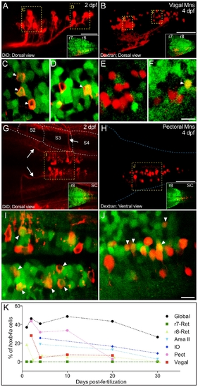
Hoxb4a activity in vagal and r8-pectoral motoneurons. (A–B, G–H) Composite dorsal views from 125 μm (A), 180 μm (B), 130 μm (G) and 70 μm (H) confocal stacks showing retrogradely labeled vagal (A–B) and pectoral (G–H) motoneurons at 2 and 4 dpf. Insets show the corresponding hoxb4a expression. (C–F, I–J) Single plane high magnification images showing hoxb4a-YFP expression and retrogradely labeled motoneurons. Arrows point to co-labeled cells. Anterior is to the left. Scale bars = 50 μm (A–B, G–H) and 10 μm (C–F, I–J). (K) Percentage of hoxb4a cells in each identified neuronal subgroup versus time from 2–30 dpf. |
Expression Data
Expression Detail
Antibody Labeling
Phenotype Data
Phenotype Detail
Acknowledgments
This image is the copyrighted work of the attributed author or publisher, and
ZFIN has permission only to display this image to its users.
Additional permissions should be obtained from the applicable author or publisher of the image.
Full text @ PLoS One