
|
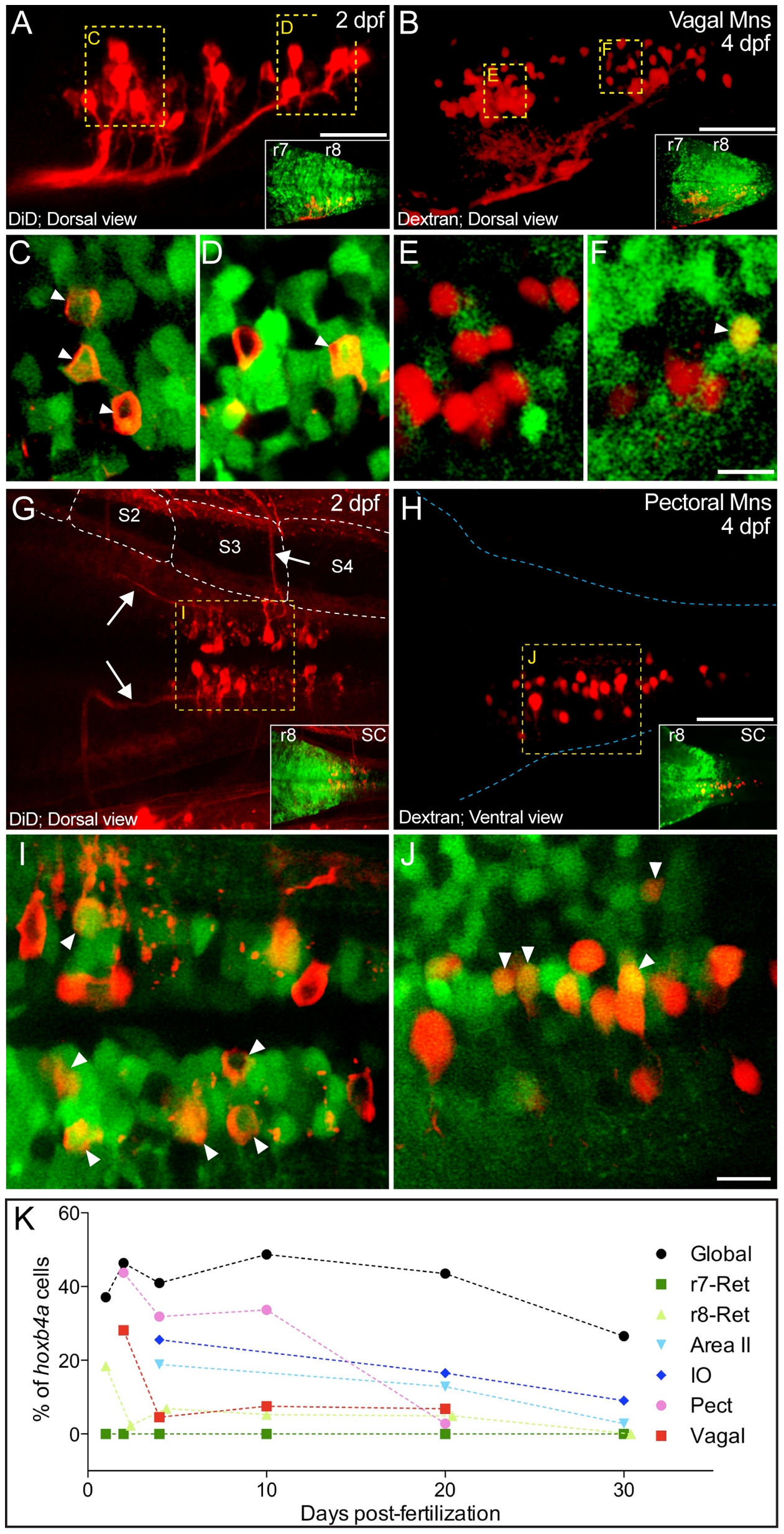
Fig. 7 Hoxb4a activity in vagal and r8-pectoral motoneurons.
(A–B, G–H) Composite dorsal views from 125 μm (A), 180 μm (B), 130 μm (G) and 70 μm (H) confocal stacks showing retrogradely labeled vagal (A–B) and pectoral (G–H) motoneurons at 2 and 4 dpf. Insets show the corresponding hoxb4a expression. (C–F, I–J) Single plane high magnification images showing hoxb4a-YFP expression and retrogradely labeled motoneurons. Arrows point to co-labeled cells. Anterior is to the left. Scale bars = 50 μm (A–B, G–H) and 10 μm (C–F, I–J). (K) Percentage of hoxb4a cells in each identified neuronal subgroup versus time from 2–30 dpf.