
Fig. 3
- ID
- ZDB-FIG-090617-32
- Publication
- Bae et al., 2009 - Anatomy of zebrafish cerebellum and screen for mutations affecting its development
- Other Figures
- All Figure Page
- Back to All Figure Page
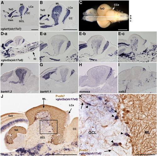
Glutamatergic neurons. (A, B) Expression of vglut1 (slc17a7) in the adult cerebellum. In situ hybridization of a sagittal section. Low (A) and high (B) magnification views. Anterior is to the left. vglut1 expression is detected in the torus longitudinalis (TL) of the TeO and the GCL of the Va, CCe, and LCa. The vglut1+ region between TeO and GCL (indicated by arrowhead) belongs to the GCL (Suppl. Fig. 1). (C, D, E) Expression of vglut2a (slc17a6). Sagittal sections at the levels (a medial, b mediolateral, c lateral.) shown in (C) were stained with a vglut2a (slc17a6) riboprobe. Low (D-a) and high (E-a, E-b, E-c) magnification views. vglut2ahigh cells were scarcely observed in the medial region (D-a), but many were detected in more lateral regions (E-b, E-c). Expression of vglut2a (slc17a6) in the inferior olive nucleus is marked by an arrow (D-a). (F–I) Differential expression of barhl1.2 (F), barhl1.1 (G), eomesodermin a (eomesa, H), and calbindin2 (calb2, I). (J, K) vglut2ahigh eurydendroid cells. Co-staining with a vglut2a riboprobe (purple, NBT/BCIP staining) and anti-Pvalb7 antibody (brown, DAB staining). (K) High-magnification view of the box in J. Note that vglut2ahigh cells (arrowheads) receive inputs from the Pvalb7+ axons of Purkinje cells (arrows). The abbreviations are described in Fig. 1. Scale bars: 500 μm (A, B, C, J) and 100 μm (K). The magnification of D and E–I are the same as in A and B, respectively. |
| Genes: | |
|---|---|
| Fish: | |
| Anatomical Terms: | |
| Stage: | Adult |
Reprinted from Developmental Biology, 330(2), Bae, Y.K., Kani, S., Shimizu, T., Tanabe, K., Nojima, H., Kimura, Y., Higashijima, S.I., and Hibi, M., Anatomy of zebrafish cerebellum and screen for mutations affecting its development, 406-426, Copyright (2009) with permission from Elsevier. Full text @ Dev. Biol.