
Fig. 12
- ID
- ZDB-FIG-090617-41
- Publication
- Bae et al., 2009 - Anatomy of zebrafish cerebellum and screen for mutations affecting its development
- Other Figures
- All Figure Page
- Back to All Figure Page
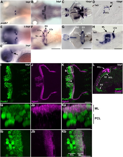
Development of the cerebellum at larval stages. (A–D) Expression of parvalbumin7 (pvalb7) at 3 dpf (A), 5 dpf (B), 10 dpf (C), and 15 dpf (D). (E–H) Expression of vglut1(slc17a7) at 3.5 dpf (E), 5 dpf (F), 10 dpf (G), and 15 dpf (H). Dorsal views (A–C, E–G), lateral view at 3 dpf (E′), and sagittal sections (D, H). (I–K) Co-staining of 5-dpf larvae with anti-Pvalb7 (green) and anti-Vglut1 (magenta) antibodies. Pvalb7 (I, Ia, Ib) and Vglut1 (J, Ja, Jb) signals and merged images (K, Ka, Kb) are shown. (I–K) Dorsal views with anterior to the left. (Ia–Ka) Transverse-section images of box a in K generated by Z-stack data, with dorsal to the top. (Ib–Kb) High-magnification view of box b in K, with anterior to the left. Pvalb7+ dendrites extended to the ML dorsally (Ka) and posteriorly (Kb), and interacted with Vglut1+ presynaptic terminals of the parallel fibers at 5 dpf. (L) Co-staining 5-dpf cerebellum with a vglut1 riboprobe (green) and anti-Pvalb7 antibody (magenta). Transverse section with dorsal to the top. The ventricular zone (VZ) is indicated by a dotted line. The vglut1+ granule cell layer (GCL) formed beneath the Purkinje cell layer (PCL) by 5 dpf. EG, eminentia granularis; other abbreviation are described in Fig. 1. Scale bars: 500 μm (G), 200 μm (F), 100 μm (H), and 50 μm (K, Ka, Kb, L). |
Reprinted from Developmental Biology, 330(2), Bae, Y.K., Kani, S., Shimizu, T., Tanabe, K., Nojima, H., Kimura, Y., Higashijima, S.I., and Hibi, M., Anatomy of zebrafish cerebellum and screen for mutations affecting its development, 406-426, Copyright (2009) with permission from Elsevier. Full text @ Dev. Biol.