
Fig. 2
- ID
- ZDB-FIG-090520-9
- Publication
- Thisse et al., 1994 - Goosecoid expression in neurectoderm and mesendoderm is disrupted in zebrafish cyclops gastrulas
- Other Figures
- All Figure Page
- Back to All Figure Page
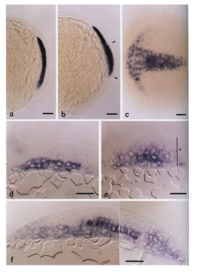
Localization of gsc transcripts by in situ hybridization during gastrulation in wild-type embryos. (a) Whole mount at 70% epiboly in lateral view. gsc RNA is restricted to axial mesendoderm (animal pole up; dorsal, right). (b) Lateral view of a whole mount embryo at 80% epiboly showing both mesendodermal and ectodermal (between the arrowheads) gsc expression (animal pole up; dorsal, right). (c) Animal pole view at the end of gastrulation, focusing on the new anterolateral expression of gsc (anterior, left). (d) Transverse section at 75% epiboly. Abundant gsc transcripts are detected in mesendoderm and smaller quantities are observed for the first time in ectodermal cells in contact with axial mesendoderm (dorsal, top; ventral, bottom). (e) Transverse section at 90% epiboly. Two gradients of gsc transcripts are detected in ectoderm: one along ventrodorsal and the other along mediolateral coordinates, both decreasing from a maximum in the ventral midline ectoderm. (f) Sagittal section at the end of gastrulation. In the mesendoderm, staining extends from the anterior tip of the embryo, including the presumptive hatching gland (left) to the middle of the gsc-expressing ectodermal territory. In ectoderm, the anterior labeling on top of the presumptive hatching gland is continuous with the anterolateral domain shown in (b). Note the intense labeling in axial ectodermal cells directly in contact with the axial mesendoderm. This labeling decreases in neighboring cells dorsally and posteriorly (anterior, left; dorsal, top). Scale bars, 100 μm (a-c), 40 μm (d-f). |
Reprinted from Developmental Biology, 164, Thisse, C., Thisse, B., Halpern, M.E., and Postlethwait, J.H., Goosecoid expression in neurectoderm and mesendoderm is disrupted in zebrafish cyclops gastrulas, 420-429, Copyright (1994) with permission from Elsevier. Full text @ Dev. Biol.