
Fig. 4
- ID
- ZDB-FIG-090520-11
- Publication
- Thisse et al., 1994 - Goosecoid expression in neurectoderm and mesendoderm is disrupted in zebrafish cyclops gastrulas
- Other Figures
- All Figure Page
- Back to All Figure Page
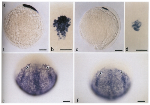
Extent of cephalic mesendodermal alteration in cycbl6 mutant embryos. (a) Side view and (b) dorsal view of the same wild-type embryo at 100 % epiboly labeled with hatching gland gene 1 (hggl) RNA probe. (c) Side view and (d) dorsal view of a cyclops embryo from the same in situ hybridization reaction (24.1% mutants = 36/149), showing strong reduction of the hatching gland territory. (e) Animal pole view of a wild-type embryo at 100% epiboly labeled with snail2 RNA probe, snail2 RNA is detected in neural crest cells, in a subset Of paraxial and lateral cephalic mesoderm, and in the prechordal plate caudal to the hatching gland territory (unpublished data). In wild type, the axial mesendoderma! staining is continuous and easily distinguishable from the surrounding loose network of paraxial and lateral mesendodermal label. (f) Animal pole view of a cyclops embryo (22.4% mutants = 11/49). Labeling of the prechordal plate is greatly reduced and apparently replaced by staining characteristic of snail2 in paraxial mesendoderm. As an internal control, neural crest cell labeling (arrowheads) appears normal in the cyclops embryo. Scale bars, 100 μm (a and c; dorsal on right; b, d-f, anterior on top). |
Reprinted from Developmental Biology, 164, Thisse, C., Thisse, B., Halpern, M.E., and Postlethwait, J.H., Goosecoid expression in neurectoderm and mesendoderm is disrupted in zebrafish cyclops gastrulas, 420-429, Copyright (1994) with permission from Elsevier. Full text @ Dev. Biol.