
Fig. 5
- ID
- ZDB-FIG-090513-39
- Publication
- Cooper et al., 1996 - A cluster of noninvoluting endocytic cells at the margin of the zebrafish blastoderm marks the site of embryonic shield formation
- Other Figures
- All Figure Page
- Back to All Figure Page
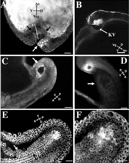
Location of forerunner cells in tail rudiment. Scale bars, 50 μm. Embryos in A, E, and F were colabeled with SYTO-11 and BODIPY 505/515 at dome stage. The embryos in B, C, and D were labeled only with SYTO-11. (A) The forerunner cell cluster with brightly stained nuclei (white arrow) in a tailbud-stage embryo is located ventral to the chordoneural hinge and germ ring on the dorsal side of the embryo. A portion of the forerunner cell cluster forms the dorsal roof of Kupffer’s vesicle (KV), the dark fluid-filled compartment. The YSL forms the ventral surface of the vesicle. The compacted YSL presses toward the animal pole into the central mass of yolk platelets within the embryo’s yolk cell (the edge of the YSL is delineated by black arrows). The forerunner cell cluster in this embryo was imaged from 70%-epiboly to blastopore closure (see Figs. 4D-4F). (B) In a tailbud-stage embryo, the forerunner cells form the dorsal roof of Kupffer’s vesicle (KV) (arrow). Side view of embryo. (C) A different embryo at the 16- to 18-somite stage with a side view of Kupffer’s vesicle surrounded by the brightly labeled (white arrow) forerunner cells. Kupffer’s vesicle is now located on the ventral side of the tail rudiment. (D) Localization of a forerunner cell cluster in an embryo at the approximately 20-somite stage. The forerunner cells surround Kupffer’s vesicle. The faintly visible yolk tube (arrow) is located on the ventral side of the extending tail. The embryo is viewed from the side, with the dorsal side of the tail facing to the top right. (E) The forerunner cell cluster remains ventral to the chordoneural hinge during early tail extension. The embryo is at approximately the 26-somite stage. Kupffer’s vesicle has disappeared by this stage of development. Side view of tail rudiment with its dorsal side facing down. (F) The same embryo as in E at a higher magnification and at a more lateral plane of focus (notochord no longer visible). Labeled cells have detached from the main forerunner cell mass. |
Reprinted from Developmental Biology, 180(1), Cooper, M.S. and D'Amico, L.A., A cluster of noninvoluting endocytic cells at the margin of the zebrafish blastoderm marks the site of embryonic shield formation, 184-198, Copyright (1996) with permission from Elsevier. Full text @ Dev. Biol.