
Fig. 7
- ID
- ZDB-FIG-090513-37
- Publication
- Cooper et al., 1996 - A cluster of noninvoluting endocytic cells at the margin of the zebrafish blastoderm marks the site of embryonic shield formation
- Other Figures
- All Figure Page
- Back to All Figure Page
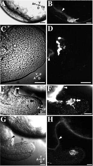
Some NEM cells are in cytoplasmic confluence with the YSL in late-blastula-stage embryos. The YSL in each embryo was loaded with Texas Red-dextran (3000 MW) by impalement loading at the oblong stage of development. The embryos were then observed at later development stages. B, D, F, and H show different results obtained by these impalement experiments. A diffuse distribution of fluorescent dextran in a forerunner cell indicates that the cell was in cytoplasmic confluence with the yolk cell at the time of impalement loading. Vesicular labeling indicates that the membrane-impermeant dextran was internalized by endocytosis. Most embryos exhibited a mixture of these two staining patterns in the forerunner cells. Corresponding Nomarski (left column) and fluorescence (right column) images are shown for each embryo. Scale bar, 50 μm. (A) Nomarski image of an approximately 75%-epiboly-stage embryo at the dorsal margin. The embryo is viewed from the side with its dorsal aspect facing down. The black arrow and arrowhead point to the locations of the forerunner cell cluster and YSL respectively, and correspond to those locations seen in B. (B) Some forerunner cells (arrow) are diffusely labeled with fluorescent dextran. Note the bright, diffusely labeled YSL (arrowhead). (C) An embryo’s tail viewed at the 14-somite stage from the side. The dorsal aspect of the tail is toward the bottom of the image. The most posterior part of the curved tail is marked by an asterisk (*). The black arrowhead points to the YSL and posterior part of the yolk tube and corresponds to the arrowhead in D. (D) This image shows an embryo with diffuse-dextran labeling in only a few of the forerunner cells. (E) Nomarski image of a tail of a 16-somite-stage embryo viewed from the side. The arrows correspond to the location of the arrows shown in F. (F) Forerunner cells exhibit both diffuse (thin arrow) and vesicular (thick arrow) fluorescent – dextran labeling. Forerunner cells are no longer in contact with the YSL (white arrowhead). (G) Nomarski image of a 16-somite-stage embryo in the same orientation as E and F but at a lower magnification. (H) The forerunner cells in this embryo show prominent vesicular labeling. Diffuse dextran labeling is present in the YSL (white arrowhead), which extends into the yolk tube at the base of the tail rudiment. |
Reprinted from Developmental Biology, 180(1), Cooper, M.S. and D'Amico, L.A., A cluster of noninvoluting endocytic cells at the margin of the zebrafish blastoderm marks the site of embryonic shield formation, 184-198, Copyright (1996) with permission from Elsevier. Full text @ Dev. Biol.