
Fig. 2
- ID
- ZDB-FIG-090513-33
- Publication
- Cooper et al., 1996 - A cluster of noninvoluting endocytic cells at the margin of the zebrafish blastoderm marks the site of embryonic shield formation
- Other Figures
- All Figure Page
- Back to All Figure Page
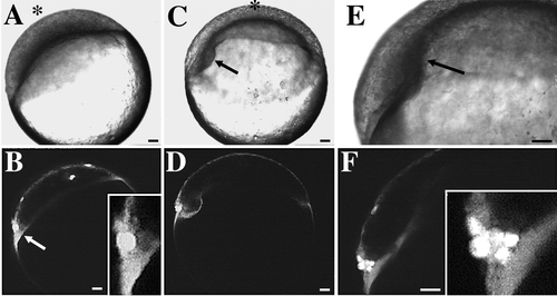
Noninvoluting endocytic marginal (NEM) cells in a late-blastula-stage embryo predict the site of embryonic shield formation. Scale bars, 50 μm. A–F show the development of an embryo previously labeled at dome stage with SYTO-11. Correlated Nomarski and confocal fluorescence micrographs at three different time points are shown. The animal pole is denoted by an asterisk. (A, B) 30%-epiboly; initial observation, 0:00 hr. (A) The blastoderm has begun to spread over the surface of the underlying yolk cell. (B) Bright cells (arrow) at the left margin of the blastoderm predict the location where the embryonic shield will form at 50%- to 60%-epiboly (see C and D). Inset (higher magnification) shows that these cells are in contact with the YSL. Several individual EVL cells that are scattered over the surface of the blastoderm are also labeled with SYTO-11. (C, D) 55%-epiboly; 1:30 hr. (C) The nascent embryonic shield is now visible as a thickening on the left side of the blastoderm (arrow). The embryo has rolled slightly into a new orientation. The animal pole now is at the top of the image. (D) The location of the fluorescently labeled NEM cell cluster in B predicted the site of embryonic shield formation. (E, F) 65%-epiboly; 2:10 hr. (E) Embryonic shield (arrow) extends toward animal pole. (F) During germ ring and embryonic shield formation, NEM cells remain at the dorsal margin of the blastoderm. NEM cells do not undergo involution during this time period. More cells are now visible in the NEM cell cluster. During embryonic shield formation, some deep cells in the NEM cell cluster move in the front of the dorsal blastoderm, at which point they become known as ‘‘forerunner’’ cells. Inset (higher magnification) shows that some forerunner cells are in contact with the YSL. |
Reprinted from Developmental Biology, 180(1), Cooper, M.S. and D'Amico, L.A., A cluster of noninvoluting endocytic cells at the margin of the zebrafish blastoderm marks the site of embryonic shield formation, 184-198, Copyright (1996) with permission from Elsevier. Full text @ Dev. Biol.