
FIGURE
Fig. 5
- ID
- ZDB-FIG-090506-28
- Publication
- Laguerre et al., 2009 - Mitotic patterns in the migrating lateral line cells of zebrafish embryos
- Other Figures
- All Figure Page
- Back to All Figure Page
Fig. 5
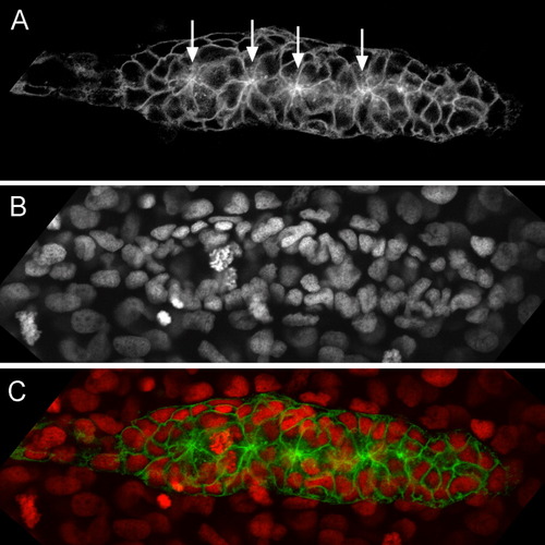
Rosette formation. A: The apical constriction of radially organized cells forming a rosette is easily detected in the Tg(-8.0cldnb:lynGFP) line (arrows). B: Rosette organization is also evidenced by the position of the nuclei, which are deeper at the center of the rosette than in its annulus; the nuclei are labeled with Hoechst. C: Superimposition of A and B falsely colored, to illustrate the correspondence between apical spots and deep nuclei. This primordium is unusual in having four rosettes instead of the usual three. |
Expression Data
Expression Detail
Antibody Labeling
Phenotype Data
Phenotype Detail
Acknowledgments
This image is the copyrighted work of the attributed author or publisher, and
ZFIN has permission only to display this image to its users.
Additional permissions should be obtained from the applicable author or publisher of the image.
Full text @ Dev. Dyn.