|
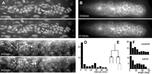
Unpaired and paired mitoses in green fluorescent protein (GFP) reporter lines. A: Metaphase (arrow) and telophases (arrows) visualized in the Tg(h2afv:GFP) line. B: Premitotic cell (arrow) and postmitotic cells (arrows) visualized in the Tg(-8.0cldnb:lynGFP) line; note that the adjacent cell (arrowhead) has a typically premitotic morphology. C: Unpaired mitosis (arrowhead) and paired mitoses (arrows) in the Tg(h2afv:GFP) line. D: Distribution of time intervals between adjacent mitoses. E: Schematic drawing of the proposed determinate lineage. The two paired mitoses are represented by the black circle; the unpaired mitoses by white ones. Plain lines: four-cell lineage; dashed lines: five-cell lineage (see text). F: Distribution of the angles between axes of division and of migration for unpaired and paired mitoses.
|