
Fig. 4
- ID
- ZDB-FIG-090504-34
- Publication
- Kalén et al., 2009 - Combination of reverse and chemical genetic screens reveals angiogenesis inhibitors and targets
- Other Figures
- All Figure Page
- Back to All Figure Page
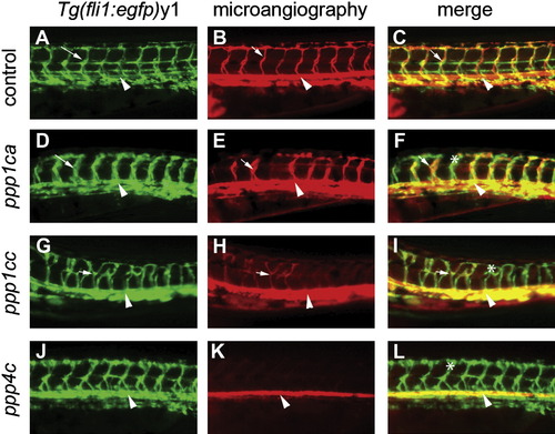
Knockdown of ppp1ca, ppp1cc, and ppp4c Results in Defects in Endothelial Path Finding and Tubulogenesis (A–L) Images are all lateral views of the trunk vasculature at 48–50 hpf. (A–C) Control embryo injected with a mixed-base morpholino. (D–L) Embryos injected with morpholinos against (D–F) ppp1ca, (G–I) ppp1cc, and (J–L) ppp4c. Endothelial cells (shown with green fluorescence in [A], [D], [G], and [J]) were identified by using the Tg(fli1:egfp)y1 line. Dorsal aortas are marked with arrowheads, examples of perfused ISVs are marked with arrows, and examples of nonperfused ISVs are labeled with asterisks. (D and J) The ppp1ca and ppp4c knockdowns resulted in enlarged ISVs, whereas (G) knockdown of ppp1cc resulted in excessive branching of the ISVs. (B) At 48–50 hpf, circulation as observed by microangiography was observed in control injected embryos (mixed-base MPO) in the dorsal aorta, cardinal vein, and ISVs. (E and K) Rhodamine-dextran dye (red) often entered the ventral aspect of the ISVs in the ppp1ca and ppp4c knockdowns (a more severely affected embryo is shown for ppp4c), but a circulatory loop was not established. (H) The ppp1cc knockdown embryos showed either an absence of circulation or thin vessels with reduced circulation. (C, F, I, and L) Merged images of the embryos shown in the previous two panels. |
| Fish: | |
|---|---|
| Knockdown Reagents: | |
| Observed In: | |
| Stage: | Long-pec |