
Fig. 4
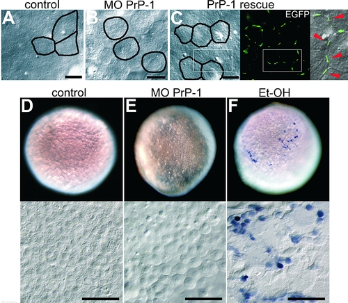
Effect of PrP-1 Knockdown in Embryonic Cell Adhesion Differences in tissue compactness between deep cells of control, morphant, and rescued embryos were evaluated at the shield stage (6 hpf ). (A) Control embryos exhibit normal tissue compactness and polygonal cell shapes. (B) Reduced cell adhesion and rounded cells are evident in PrP-1 morphant (MO) embryos. (C) Local accumulation of EGFP-PrP-1 at cell contacts (see red arrowheads in detailed overlay view of framed region, right) reverts these effects in rescued embryos. Cell outlines were digitally redrawn to help visualization of cell shape. (D–F) The loss of embryonic cell adhesion is not related to cell-death, as whole-mount TUNEL stainings of control (D), morphant (E), and ethanol-treated embryos (F) show apoptotic cells (blue staining) only in (F). Scale bars in (A–C) indicate 10 μm; scale bars in (D–F) indicate 50 μm. |
| Fish: | |
|---|---|
| Knockdown Reagents: | |
| Observed In: | |
| Stage: | Shield |