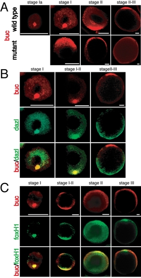
buc, dazl, and foxH1 mRNA Localization during Oogenesis
Whole-mount in situ hybridization with buc (red) and dazl mRNA (green) or foxH1 mRNA (green). Animal to the top. Some of these panels are double stainings of the single channels displayed in Fig. 2 to confirm the localization of buc mRNA.
(A) In wild type oocytes buc mRNA is localized at the initiation of oogenesis (stage IA) in the Balbiani body, until stage II at the vegetal pole and finally during the transition from stage II to III at the animal pole. In buc mutants buc mRNA is localized at the animal pole until stage II and disappears between stages II and III. Note that the mutant oocyte at stage II-III is three times longer exposed than at stage I.
(B) Until stage II buc and dazl mRNA colocalize (yellow in merged picture, lower row). Then dazl mRNA remains vegetal, whereas buc is detected animal (last column).
(C) During all stages buc and foxH1 mRNA co-localize (yellow in merged pictures, lower row). Scale bar: 25 μm (stage I), 50 μm (all other stages).
|
