
Fig. 4
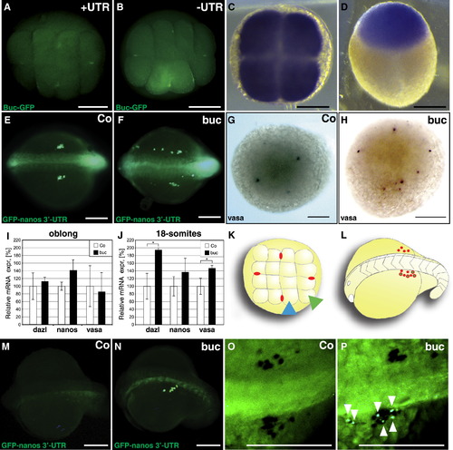
Buc Colocalizes with Germ Plasm and Induces the Formation of Germ Cells during Embryogenesis (A and B) Buc-GFP localizes to the germ plasm in eight-cell embryos. One-cell embryos were injected with 200 pg mRNA-encoding Buc-GFP (green) with (A) or without 3′UTR (B). Note the localization of GFP at four cleavage furrows in living eight-cell embryos. Animal view. Scale bars represent 200 μm. (C and D) buc mRNA is throughout the blastodisc during embryogenesis. Whole-mount in situ hybridization showing buc mRNA in the blastodisc (blue) without specific localization to the germ plasm at the cleavage furrow at four-cell (C) and blastula stages (D) (high stage). Animal view (C), lateral view, animal to the top (D). Scale bars represent 200 μm. (E–J) Overexpression of Buc in one-cell embryos induces germ cell formation. Dorsal view of living 13 somite stage embryos (15.5 hpf), anterior to the left (E and F). Germ cells are labeled green fluorescent after coinjection with 100 pg GFP-nanos-3′UTR mRNA. In control embryos, germ cells accumulate in the bilateral gonad anlagen (E), whereas in buc-injected embryos, extragonadal germ cells are visible (F). Animal view of oblong stage embryos (3.5 hpf) after whole-mount in situ hybridization with vasa mRNA (blue) injected with 300 pg GFP (G) or buc mRNA at the one-cell stage (H). Note additional vasa-positive cells in buc-injected embryos (85.0% ± 11.5%; n = 343) compared to control injections (5.7% ± 8.7%; n = 280; ***p = 3.4 x 10⁻¹²). Scale bars represent 200 μm (E–H). (I and J) Real-time PCR analysis of the germ plasm RNAs nanos, vasa, and dazl after control and buc mRNA injection analyzed at oblong stage (3.5 hpf; [I]) and 18 somites stage (18 hpf; [J]). Error bars represent SD (dazl *p = 0.028; vasa *p = 0.041; Table S3). (K and L) Experimental scheme for germ cell formation assay. (K) The 16-cell embryos (animal view) were either injected into one corner blastomere (green arrowhead) or into a middle blastomere (blue arrowhead; positive control) containing essential germ plasm (red ovals). (L) Embryos were examined between the 13 and 18 somite stage for the formation of additional germ cells (green dots) in addition to the endogenous germ cells (red dots; not visible in the experiment). Oblique dorsal view, anterior to the left. (M and N) Live 15 somite stage embryos, similar view as in panel (L), after injection of 100 pg GFP-nanos 3′-UTR mRNA into a corner blastomere (M) or after coinjection of 170 pg buc mRNA into the corner blastomere (N) with 12 ± 5.4 (n = 21) fluorescent cells per embryo. Scale bars represent 200 μm. (O and P) Buc-induced germ cells express vasa mRNA. Injection of GFP-nanos 3′-UTR into a corner blastomere of a 16-cell embryo does not label germ cells after vasa mRNA in situ hybridization at the 18 somite stage (black; [O]), whereas coinjection of buc mRNA generates additional vasa-positive cells also expressing GFP (green, white arrowheads; [P]). Dorsal view, anterior to the left. Scale bars represent 200 μm. |
| Gene: | |
|---|---|
| Fish: | |
| Anatomical Term: | |
| Stage Range: | 4-cell to High |