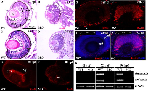
A-D: The histological examination of zebrafish retinae. Histological section and hematoxylin-eosin staining of retinae were performed on wild-type embryos and arl6ip-morphants at 72 and 96 hpf. Unlike the wild-type embryos (A,C), the neuronal differentiation is severely delayed, resulting in no formation of retinal layers in the arl6ip-morphants, both at 72 (B) and 96 hpf (D). In addition, retina development of arl6ip-morphants was markedly retarded. GCL, ganglion cell layer; INL, inner nuclear layer; L, lens; MO, arl6ip-morphant; ONL, outer nuclear layer; ON, optic nerve; OPL, outer plexiform layer; IPL, inner plexiform layer; PCL, pigment cell layer; WT, wild-type. The scale bar = 100 μm. E,F: Knockdown of Arl6ip causes the defective formation of the retinal axons in zebrafish embryos. By using the confocal fluorescence microscope, we observed that Zn8 protein was detected in the retinal ganglion cell axons of wild-type (WT) embryos at 48 hpf (E). However, the protein level of Zn8 presented a very weak signal near the retinal ventral (F, arrow) in the embryos injected with 4 ng/embryo of arl6ip-MO1 (MO). GCL, ganglion cell layer; ON, optic nerve. G-J: The retinal cells stayed at S-phase in retinal developmental period in zebrafish embryos when Arl6ip was lost. BrdU (red) and DAPI (blue) were used to label the S-phase of cell cycle and the nuclear, respectively. There was no BrdU signal detected in the outer nuclear layer (onl), inner nuclear layer (inl), and ganglion cell layer (gcl) (G,I) of wild-type embryos (WT) at 72 hpf, except the ciliary marginal zone (cmz, arrowhead). However, at the same stage, in embryos injected with 4 ng/embryo of arl6ip-MO (MO), we found that there was a large number of BrdU signal in retina (H,J), indicating that the retina cells of arl6ip-morphant were maintained at S-phase. The images were taken by confocal microscopy. The scale bars = 75 μm (G,I) and 50 μm (H,J). K: Detection of the transcripts of rhodopsin and red opsin mRNA of zebrafish embryos using RT-PCR. After the total RNA was extracted from wild-type embryos and arl6ip-morphants at 48, 72, and 96 hpf, RT-PCR was used to detect the existence of the transcripts of rhodopsin, which are expressed exclusively in rods and red cones, respectively. The expression of tubulin served as positive control. Results showed that neither rhodopsin nor red opsin transcripts were detected in either the wild-type embryos or arl6ip-morphants at 48 hpf as a result of the fact that the opsin gene is not transcribed until 50 hpf in the retina. After starting transcription of rhodopsin mRNA and red opsin mRNA at, for example, 72 and 96 hpf, the transcripts of rhodopsin and red opsin were present in the wild-type embryos. However, both rhodopsin and red opsin mRNA were totally absent in the arl6ip-morphants. WT, wild-type; MO, arl6ip-morphants.
|
