
Fig. 4
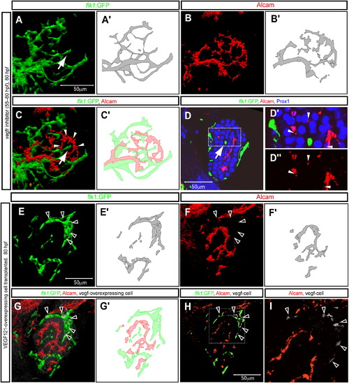
Endothelial Cells Can Influence Alcam Subcellular Localization (A–D) Tg(flk1:EGFP)s843 larvae treated with the VEGF-receptor inhibitor SU5416 from 55 to 80 hpf were visualized for GFP (green), Alcam (red), and Prox1 (blue) expression at 80 hpf. (A) Projected confocal image of Tg(flk1:EGFP)s843 expression in the liver of an SU5416-treated larva in which the intrahepatic vascular network (schematically presented in [A′]) is disrupted and endothelial cells have formed a dead-end vascular branch (indicated by arrow). (B) Projected confocal image of the same liver visualized for Alcam expression at 80 hpf. The pattern of the Alcam-positive network (schematically presented in [B′]) reflects the disrupted intrahepatic vascular network. (C) Merged image of Alcam (red) and Tg(flk1:EGFP)s843 (green) expression shows that the intrahepatic networks still do not intersect. (D) Z-plane confocal image of the same larva. Alcam (indicated by white arrowheads) accumulates on membranes away from the dead-end vascular branch (indicated by arrow). The outlined area is magnified and shown in (D′). Alcam immunostaining in (D′) is shown separately in (D″). (E–I) Tg(flk1:EGFP)s843 larva containing vegf121 mRNA-overexpressing cells visualized for GFP (green), Alcam (pseudocolored red), and donor cell tracer (rhodamine dextran; pseudocolored white) at 80 hpf. (E) Projected confocal image of Tg(flk1:EGFP)s843 expression in the liver. The transplanted vegf121 mRNA-overexpressing cells (indicated by black arrowheads) disrupt the intrahepatic vascular network (schematically presented in [E′]). (F) Projected confocal image of the same liver visualized for Alcam expression at 80 hpf. The pattern of the Alcam-positive network (schematically presented in [F′]) reflects the disrupted intrahepatic vascular network. (G) Merged image of Alcam (red) and Tg(flk1:EGFP)s843 (green) expression shows that the intrahepatic vascular networks still do not intersect. (H) Z-plane confocal image of the same larva. The outlined area is magnified and shown in (I). |
| Genes: | |
|---|---|
| Antibodies: | |
| Fish: | |
| Condition: | |
| Anatomical Terms: | |
| Stage: | Protruding-mouth |