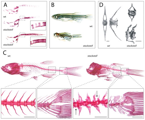
stocksteif mutants exhibit over-ossification of the larval and adult vertebral column. (A) Alizarin Red bone staining of a wild-type embryo and two stocksteif embryos at 8 dpf. Insets show that the initial spacing of vertebrae is normal (upper inset), but that excessive bone formation causes fusion of the future vertebrae (both insets). (B) Sibling wild-type and stocksteif juveniles (2 months). Pictures are taken at the same magnification. (C) Alizarin Red bone staining of wild-type and stocksteif juveniles (2 months) shown to scale with magnifications of part of the vertebral column and the tail. In mutants, vertebrae are usually fused with each other, and only few intervertebral boundaries can be observed (arrows). Hypural elements in the tail are also fused. Note expansion of both neural and haemal arches in mutants. Scale bars: 500 μm. (D) MicroCT scans of wild-type (3 months) and stocksteif juveniles (2 months) show that mutants exhibit completely solid centra, with the central opening (area of the nucleus pulposus) completely filled by excess bone. Scale bars: 100 μm.
|
