
Fig. 2
- ID
- ZDB-FIG-081028-12
- Publication
- Ahn et al., 2008 - Tri-phasic expression of posterior Hox genes during development of pectoral fins in zebrafish: Implications for the evolution of vertebrate paired appendages
- Other Figures
- All Figure Page
- Back to All Figure Page
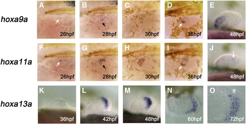
Expression of hoxa9a, hoxa11a, and hoxa13a during zebrafish pectoral fin development. Expression of hoxa9a (A–E) and hoxa11a (F–J) is largely confined to the prospective myogenic cells of the fin bud, whereas expression of hoxa13a (K–O) is seen exclusively within the distal mesenchyme cells that later give rise to the connective tissues of the fin blade. Oblique dorsal (A–D, F–I), lateral (E, J, K–M), or dorsal (N, O) views with anterior to the left in all panels. Only left side is shown for each embryo. Some embryos are also stained for muscle myosin (brown staining in panels A–D, F–I) to show the position of myotomes. Arrows in panels A/F, B/G, and D/I mark the expression within the nascent myogenic cells (A, F), myogenic cells invading the fin bud proper (B, G), and lateral cluster of prospective pectoral fin muscle cells (D, I), respectively. ff: fin fold (later, fin blade). hpf: hours post fertilization. |
| Genes: | |
|---|---|
| Antibody: | |
| Fish: | |
| Anatomical Terms: | |
| Stage Range: | Prim-5 to Protruding-mouth |
Reprinted from Developmental Biology, 322(1), Ahn, D., and Ho, R.K., Tri-phasic expression of posterior Hox genes during development of pectoral fins in zebrafish: Implications for the evolution of vertebrate paired appendages, 220-233, Copyright (2008) with permission from Elsevier. Full text @ Dev. Biol.