
Fig. 2
- ID
- ZDB-FIG-080922-12
- Publication
- Hu et al., 1999 - Retinal neurogenesis: the formation of the initial central patch of postmitotic cells
- Other Figures
- All Figure Page
- Back to All Figure Page
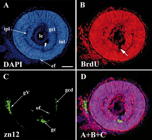
The first postmitotic retinal cells appeared at 28 hpf in ventronasal retina, and included the first ganglion cells. 28 hpf BrdU, 42 hpf sacrifice. All panels show the same (intermediate) section, stained as indicated. (A) DAPI staining shows all nuclei. (B) BrdU staining shows those (unlabeled) nuclei that had ceased DNA synthesis before 28 hpf. (C) zn12 stain shows early neurons in the retina and the trigeminal ganglion. (D) A merger of A, B, and C to show that the early ganglion cells (zn12-positive) lie in the BrdU-negative cells adjacent to the embryonic fissure. Abbreviations: as in Fig. 1, gc, ganglion cells; gcd, ganglion cell dendrites; gcl, ganglion cell layer; gV, trigeminal ganglion; inl, inner nuclear layer; ipl, inner plexiform layer; of, optic fibers. Calibration: 40 μm. |
Reprinted from Developmental Biology, 207, Hu, M. and Easter, Jr., S.S., Retinal neurogenesis: the formation of the initial central patch of postmitotic cells, 309-321, Copyright (1999) with permission from Elsevier. Full text @ Dev. Biol.