
Fig. 1
- ID
- ZDB-FIG-080922-11
- Publication
- Hu et al., 1999 - Retinal neurogenesis: the formation of the initial central patch of postmitotic cells
- Other Figures
- All Figure Page
- Back to All Figure Page
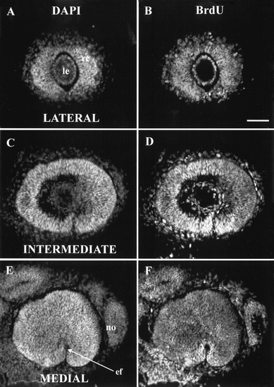
At 27 hpf, all retinal cells were synthesizing DNA. 27 hpf injection of BrdU, 41 hpf sacrifice. Three sagittal sections at the levels (lateral, intermediate, and medial) described in the text. In this and subsequent figures, all eyes are shown as right eyes viewed laterally; that is, dorsal is up and nasal (anterior) is to the right. (A, C, E) All stained with DAPI to show all nuclei; (B, D, F) all stained with anti-BrdU to show those nuclei synthesizing DNA between 27 and 41 hpf. In both the lens and the nose, some nuclei are labeled with DAPI but not BrdU, so they had ceased DNA synthesis prior to 27 hpf. No such cells were found in the retina. Abbreviations: ef, embryonic fissure; le, lens; no, nose. Calibration: 40 μm. |
Reprinted from Developmental Biology, 207, Hu, M. and Easter, Jr., S.S., Retinal neurogenesis: the formation of the initial central patch of postmitotic cells, 309-321, Copyright (1999) with permission from Elsevier. Full text @ Dev. Biol.